

Visualization of Brain Shift Corrected Functional Magnetic Resonance Imaging Data for Intraoperative Brain Mapping
- PMID: 31218295
- PMCID: PMC6580887
- DOI: 10.1016/j.wnsx.2019.100021
Visualization of Brain Shift Corrected Functional Magnetic Resonance Imaging Data for Intraoperative Brain Mapping
Abstract
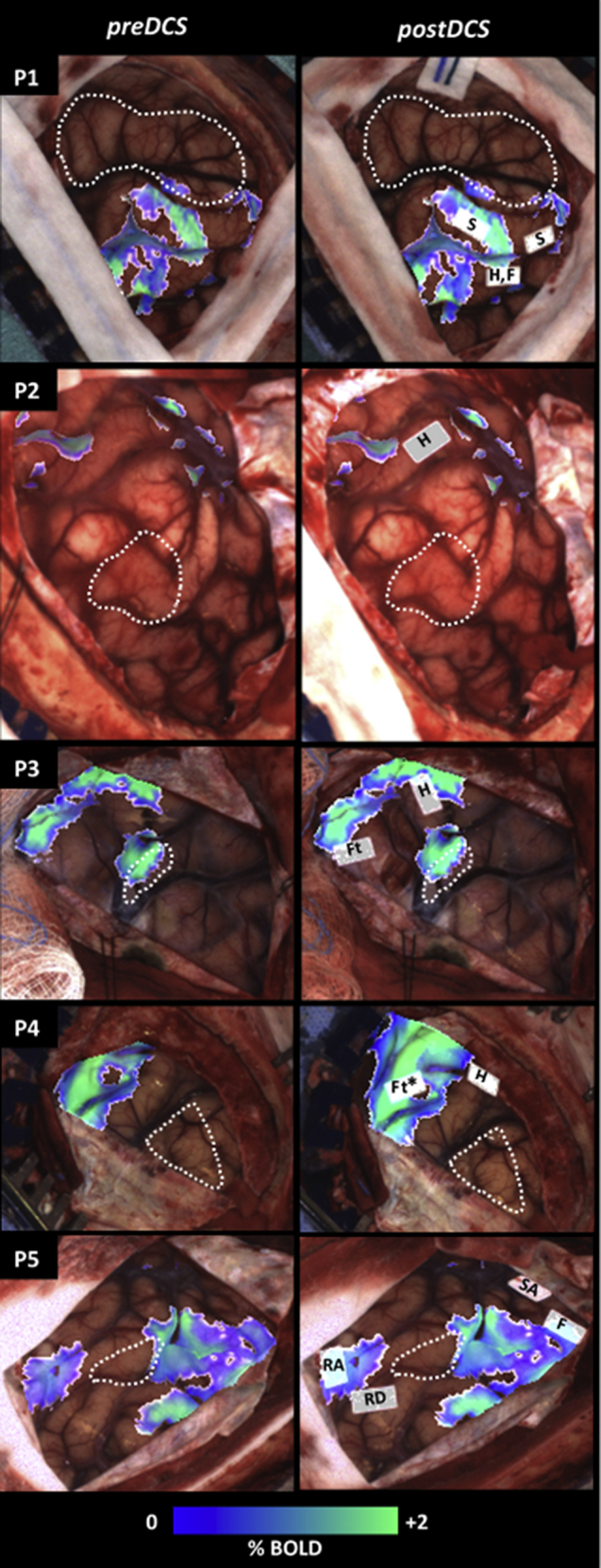
Background: Brain tumor surgery requires careful balance between maximizing tumor excision and preserving eloquent cortex. In some cases, the surgeon may opt to perform an awake craniotomy including intraoperative mapping of brain function by direct cortical stimulation (DCS) to assist in surgical decision-making. Preoperatively, functional magnetic resonance imaging (fMRI) facilitates planning by identification of eloquent brain areas, helping to guide DCS and other aspects of the surgical plan. However, brain deformation (shift) limits the usefulness of preoperative fMRI during surgery. To address this, an integrated visualization method for fMRI and DCS results is developed that is intuitive for the surgeon.
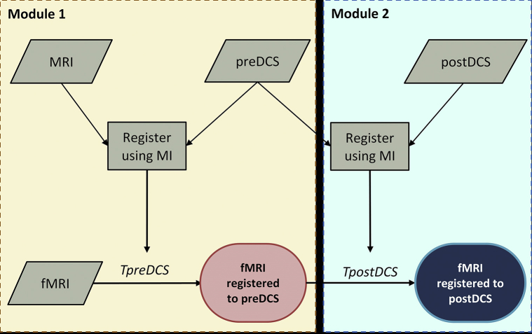
Methods: An image registration pipeline was constructed to display preoperative fMRI data corrected for brain shift overlaid on images of the exposed cortical surface at the beginning and completion of DCS mapping. Preoperative fMRI and DCS data were registered for a range of misalignments, and the residual registration errors were calculated. The pipeline was validated on imaging data from five brain tumor patients who underwent awake craniotomy.
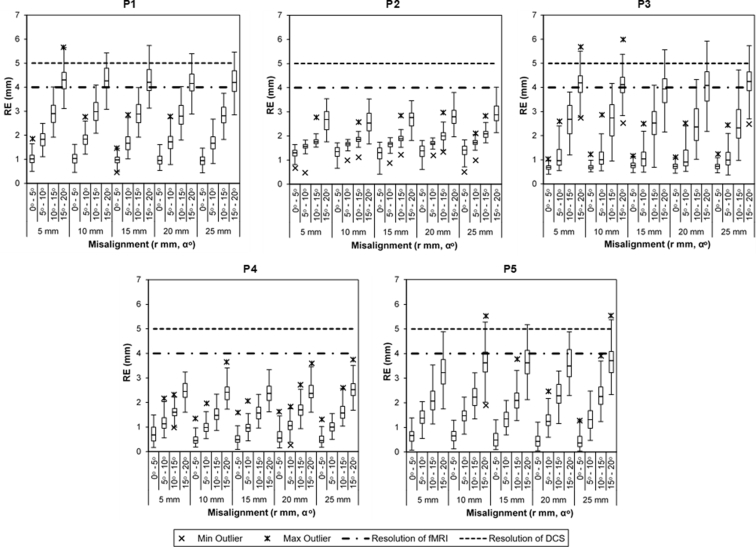
Results: Registration errors were well under 5 mm (the approximate spatial resolution of DCS) for misalignments of up to 25 mm and approximately 10-15°. For rotational misalignments up to 20°, the success rate was 95% for an error tolerance of 5 mm. Failures were negligible for rotational misalignments up to 10°. Good quality registrations were observed for all five patients.
Conclusions: A proof-of-concept image registration pipeline is presented with acceptable accuracy for intraoperative use, providing multimodality visualization with potential benefits for intraoperative brain mapping.
Keywords: 2D, 2-dimensional; 3D, 3-Dimensional; Awake craniotomy; Brain mapping; Brain tumor resection; CT, Computed tomography; DCS, Direct cortical stimulation; Electric stimulation; FOV, Field of view; Functional mapping; MRI, Magnetic resonance imaging; Multimodal imaging; RE, Registration error; Surgical planning; TE, Echo time; TR, Repetition time; fMRI, Functional magnetic resonance imaging.
Figures
Similar articles
-
Case Report: Multimodal Functional and Structural Evaluation Combining Pre-operative nTMS Mapping and Neuroimaging With Intraoperative CT-Scan and Brain Shift Correction for Brain Tumor Surgical Resection.Front Hum Neurosci. 2021 Feb 25;15:646268. doi: 10.3389/fnhum.2021.646268. eCollection 2021. Front Hum Neurosci. 2021. PMID: 33716700 Free PMC article.
-
Correlation of Preoperative Functional Magnetic Resonance Imaging (fMRI) with Intraoperative Cortical Stimulation in Surgeries of Eloquent Brain Lesions.Neurol India. 2022 Jul-Aug;70(4):1534-1539. doi: 10.4103/0028-3886.355146. Neurol India. 2022. PMID: 36076655
-
Combined noninvasive language mapping by navigated transcranial magnetic stimulation and functional MRI and its comparison with direct cortical stimulation.J Neurosurg. 2015 Jul;123(1):212-25. doi: 10.3171/2014.9.JNS14929. Epub 2015 Mar 6. J Neurosurg. 2015. PMID: 25748306
-
Is preoperative functional magnetic resonance imaging reliable for language areas mapping in brain tumor surgery? Review of language functional magnetic resonance imaging and direct cortical stimulation correlation studies.Neurosurgery. 2010 Jan;66(1):113-20. doi: 10.1227/01.NEU.0000360392.15450.C9. Neurosurgery. 2010. PMID: 19935438 Review.
-
Can Preoperative Mapping with Functional MRI Reduce Morbidity in Brain Tumor Resection? A Systematic Review and Meta-Analysis of 68 Observational Studies.Radiology. 2021 Aug;300(2):338-349. doi: 10.1148/radiol.2021204723. Epub 2021 Jun 1. Radiology. 2021. PMID: 34060940
Cited by
-
Intraoperative Brain Mapping in Multilingual Patients: What Do We Know and Where Are We Going?Brain Sci. 2022 Apr 27;12(5):560. doi: 10.3390/brainsci12050560. Brain Sci. 2022. PMID: 35624947 Free PMC article. Review.
-
An update on tests used for intraoperative monitoring of cognition during awake craniotomy.Acta Neurochir (Wien). 2024 May 7;166(1):204. doi: 10.1007/s00701-024-06062-6. Acta Neurochir (Wien). 2024. PMID: 38713405 Free PMC article.
-
Investigation of convolutional neural networks using multiple computed tomography perfusion maps to identify infarct core in acute ischemic stroke patients.J Med Imaging (Bellingham). 2021 Jan;8(1):014505. doi: 10.1117/1.JMI.8.1.014505. Epub 2021 Feb 10. J Med Imaging (Bellingham). 2021. PMID: 33585662 Free PMC article.
-
Use of a convolutional neural network to identify infarct core using computed tomography perfusion parameters.Proc SPIE Int Soc Opt Eng. 2021 Feb;11596:1159611. doi: 10.1117/12.2579753. Epub 2021 Feb 15. Proc SPIE Int Soc Opt Eng. 2021. PMID: 33707811 Free PMC article.
-
Performance of angiographic parametric imaging in locating infarct core in large vessel occlusion acute ischemic stroke patients.J Med Imaging (Bellingham). 2020 Jan;7(1):016001. doi: 10.1117/1.JMI.7.1.016001. Epub 2020 Feb 11. J Med Imaging (Bellingham). 2020. PMID: 32064301 Free PMC article.
References
-
- Petrella J.R., Shah L.M., Harris K.M. Preoperative functional MR imaging localization of language and motor areas: effect on therapeutic decision making in patients with potentially resectable brain tumors. Radiology. 2006;240:793–802. - PubMed
-
- Benson R.R., FitzGerald D.B., LeSueur L.L. Language dominance determined by whole brain functional MRI in patients with brain lesions. Neurology. 1999;52:798–809. - PubMed
-
- Sakr H.M., Mohamed M.A., Jalalod’din H., Abbas Y.A. Influence of fMRI on operative planning of brain tumors: initial experience in a histopathologically variable subset of tumors. Egypt J Radiol Nucl Med. 2011;42:215–221.
-
- Roberts D.W., Hartov A., Kennedy F.E., Miga M.I., Paulsen K.D. Intraoperative brain shift and deformation: a quantitative analysis of cortical displacement in 28 cases. Neurosurgery. 1998;43:749–758. - PubMed
LinkOut - more resources
Full Text Sources
