

Suppression of Somatosensory Evoked Cortical Responses by Noxious Stimuli
- PMID: 31218521
- PMCID: PMC6707979
- DOI: 10.1007/s10548-019-00721-z
Suppression of Somatosensory Evoked Cortical Responses by Noxious Stimuli
Abstract
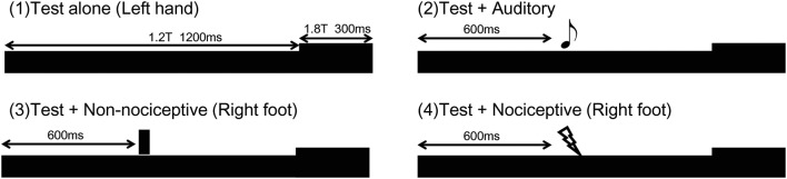
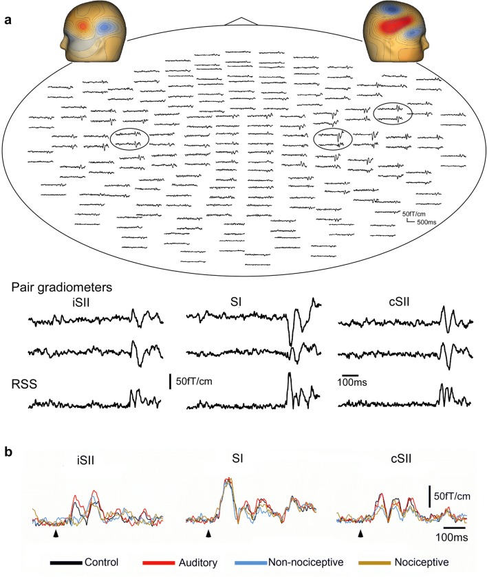
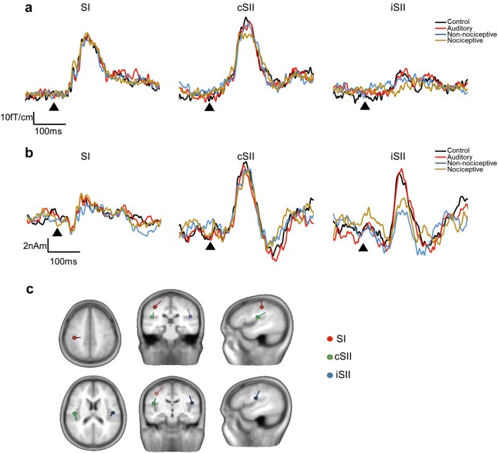
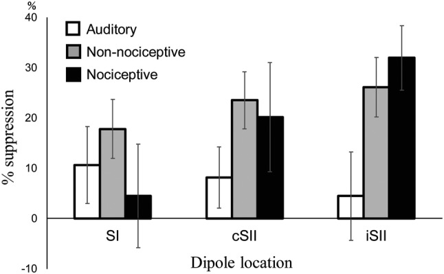
Paired-pulse suppression refers to attenuation of neural activity in response to a second stimulus and has a pivotal role in inhibition of redundant sensory inputs. Previous studies have suggested that cortical responses to a somatosensory stimulus are modulated not only by a preceding same stimulus, but also by stimulus from a different submodality. Using magnetoencephalography, we examined somatosensory suppression induced by three different conditioning stimuli. The test stimulus was a train of electrical pulses to the dorsum of the left hand at 100 Hz lasting 1500 ms. For the pulse train, the intensity of the stimulus was abruptly increased at 1200 ms. Cortical responses to the abrupt intensity change were recorded and used as the test response. Conditioning stimuli were presented at 600 ms as pure tones, either innocuous or noxious electrical stimulation to the right foot. Four stimulus conditions were used: (1) Test alone, (2) Test + auditory stimulus, (3) Test + somatosensory stimulus, and (4) Test + nociceptive stimulus. Our results showed that the amplitude of the test response was significantly smaller for conditions (3) and (4) in the secondary somatosensory cortex contralateral (cSII) and ipsilateral (iSII) to the stimulated side as compared to the response to condition (1), whereas the amplitude of the response in the primary somatosensory cortex did not differ among the conditions. The auditory stimulus did not have effects on somatosensory change-related response. These findings show that somatosensory suppression was induced by not only a conditioning stimulus of the same somatosensory submodality and the same cutaneous site to the test stimulus, but also by that of a different submodality in a remote area.
Keywords: Aδ; Change-related response; MEG; SII; Sensory gating; Sensory suppression.
Figures
Similar articles
-
Long-latency suppression of auditory and somatosensory change-related cortical responses.PLoS One. 2018 Jun 26;13(6):e0199614. doi: 10.1371/journal.pone.0199614. eCollection 2018. PLoS One. 2018. PMID: 29944700 Free PMC article.
-
Effects of prior sustained tactile stimulation on the somatosensory response to the sudden change of intensity in humans: an magnetoencephalography study.Neuroscience. 2011 May 19;182:115-24. doi: 10.1016/j.neuroscience.2011.03.019. Epub 2011 Mar 21. Neuroscience. 2011. PMID: 21420471
-
A comparative magnetoencephalographic study of cortical activations evoked by noxious and innocuous somatosensory stimulations.Neuroscience. 2003;120(1):235-48. doi: 10.1016/s0306-4522(03)00261-6. Neuroscience. 2003. PMID: 12849756
-
Inhibition of somatosensory-evoked cortical responses by a weak leading stimulus.Neuroimage. 2014 Nov 1;101:416-24. doi: 10.1016/j.neuroimage.2014.07.035. Epub 2014 Jul 24. Neuroimage. 2014. PMID: 25067817
-
The somatosensory evoked magnetic fields.Prog Neurobiol. 2000 Aug;61(5):495-523. doi: 10.1016/s0301-0082(99)00063-5. Prog Neurobiol. 2000. PMID: 10748321 Review.
Cited by
-
Assessment of haptic memory using somatosensory change-related cortical responses.Hum Brain Mapp. 2020 Dec;41(17):4892-4900. doi: 10.1002/hbm.25165. Epub 2020 Aug 26. Hum Brain Mapp. 2020. PMID: 32845051 Free PMC article.
-
Somatosensory Gating Is Modulated by Anodal Transcranial Direct Current Stimulation.Front Neurosci. 2021 Sep 7;15:651253. doi: 10.3389/fnins.2021.651253. eCollection 2021. Front Neurosci. 2021. PMID: 34557064 Free PMC article.
References
Publication types
MeSH terms
LinkOut - more resources
Full Text Sources
