. 2019 Oct;235(4):783-793.
doi: 10.1111/joa.13026. Epub 2019 Jun 20.

Hindbrain neurovascular anatomy of adult goldfish (Carassius auratus)

Affiliations

Hindbrain neurovascular anatomy of adult goldfish (Carassius auratus)

Sulman Rahmat et al. J Anat. 2019 Oct.

Abstract

The goldfish hindbrain develops from a segmented (rhombomeric) neuroepithelial scaffold, similar to other vertebrates. Motor, reticular and other neuronal groups develop in specific segmental locations within this rhombomeric framework. Teleosts are unique in possessing a segmental series of unpaired, midline central arteries that extend from the basilar artery and penetrate the pial midline of each hindbrain rhombomere (r). This study demonstrates that the rhombencephalic arterial supply of the brainstem forms in relation to the neural segments they supply. Midline central arteries penetrate the pial floor plate and branch within the neuroepithelium near the ventricular surface to form vascular trees that extend back towards the pial surface. This intramural branching pattern has not been described in any other vertebrate, with blood flow in a ventriculo-pial direction, vastly different than the pial-ventricular blood flow observed in most other vertebrates. Each central arterial stem penetrates the pial midline and ascends through the floor plate, giving off short transverse paramedian branches that extend a short distance into the adjoining basal plate to supply ventromedial areas of the brainstem, including direct supply of reticulospinal neurons. Robust r3 and r8 central arteries are significantly larger and form a more interconnected network than any of the remaining hindbrain vascular stems. The r3 arterial stem has extensive vascular branching, including specific vessels that supply the cerebellum, trigeminal motor nucleus located in r2/3 and facial motoneurons found in r6/7. Results suggest that some blood vessels may be predetermined to supply specific neuronal populations, even traveling outside of their original neurovascular territories in order to supply migrated neurons.

Keywords: brainstem; central arteries; cerebral vessels; motor nuclei; neuroepithelium; reticulospinal neurons; rhombomere; teleost.


Figures

Figure 1
Figure 1
Rhombomeric neuronal organization in adult goldfish. The segmental divisions of the hindbrain (red dashed lines) as well as the rhombomeric neuronal pattern of reticular and motor nuclei are shown in a projection of 11 consecutive parasagittal sections from one side of the brain of a specimen retrogradely labeled with dextran‐conjugated biotin from the contralateral spinal cord (black precipitate) and immunostained for choline acetyl transferase (ChAT; brown reaction product). Rhombomeres are numbered with black Arabic numerals (1–8), cranial nerve roots with black roman numerals (e.g. IIIr) and cranial motor nuclei with white (e.g. III). The ChAT staining deposits brown reaction product in cholinergic neurons; here, mostly cranial motor neurons. Reticulospinal neuronal clusters are located in the centers of each hindbrain segment. Rhombomere 8, as in most other vertebrates, comprises the entire lower medulla and may represent three or four cryptic segments. The front of the hindbrain (rhombomere 1) is just caudal to the oculomotor motor nucleus (III). The facial nerve root (VIIr) can be seen entering the brain in rhombomere 4 and projecting back to the facial motor nucleus in r6/7 (VII). III, oculomotor nucleus; IIIr, oculomotor root; IV, trochlear nucleus; IVr, trochlear root; IX, glossopharyngeal motor nucleus; IXr, glossopharyngeal root; M, Mauthner neuron; MB, midbrain; OLE, octavolateral efferent nucleus; V, trigeminal motor nucleus; VI, abducens nucleus; VII, facial motor nucleus; VIIr, facial nerve root; Vr, trigeminal root; X, vagus motor nucleus. Scale bar: 1 mm.
Figure 2
Figure 2
Overall pattern of brain arteries in teleost fishes. Lateral (A) and medial (B) views of a Salmo brain modified after Grodzinski (1946). (A) The cerebral carotid artery (CCA) divides into anterior (ACC) and posterior (PCC) cerebral carotid arteries. The PCC travels caudally and medially and fuses with the corresponding vessel from the other side to form a single midline basilar artery (BA). Prior to fusion, the PCCs give off mesencephalic arteries (Mes) and cerebellar arteries (Cer). (B) As the basilar travels down the hindbrain, multiple rhombencephalic penetrating branches, called central arteries (CAs), are given off. This posterior cerebral/basilar system of arteries is responsible for supplying the brainstem and cerebellar regions of the teleostean brain. II, optic nerve; III, oculomotor nerve; IX, glossopharyngeal nerve; V, trigeminal nerve; X, vagus nerve.
Figure 3
Figure 3
Hindbrain rhombomeric central arterial organization. The segmental patterns of midline central arterial stems (CA) from rhombomere (r) 3 to r8 are shown in single horizontal sections of the hindbrain in two different adult goldfish specimens retrogradely labeled with dextran‐conjugated biotin and counterstained with cresyl violet (B) or ChAT immunostain (A). The reticulospinal neurons (RS) are located at the center of each rhombomere (best seen in B, a bilateral spinal cord label), on either side of the midline central arterial stems. A contralateral label of dextran‐conjugated biotin followed by immunostaining with ChAT (A) shows the RS neuronal clusters, midline CA stems, and the projecting axons from the spinal cord on one side of the brain. All specimens examined showed large midbrain CAs as well as r3 and r8 stems that were significantly larger than the other hindbrain CAs. Although one large r8 stem was observed in all of these specimens, several smaller midline central arteries arise in r8 as well (A,B) in order to assist in supplying specific regions of this vast segment. Sagittal projection (C) of several dextran‐conjugated biotin‐labeled adult goldfish sections showing central arteries penetrating the midline of each rhombomere. CAs, central arterial stems; MB, midbrain; r, rhombomere; RS, reticulospinal neurons; VII mot, facial motor tract. Scale bars: 500 μm (A,B), 1 mm (C).
Figure 4
Figure 4
Paramedian branches of central arteries. Retrograde label with dextran‐conjugated biotin from the spinal cord followed by cresyl‐violet counterstain of adult goldfish brains (A–D) show the overall arterial and reticulospinal (RS) neuronal segmental pattern (A,C). The cresyl violet counterstain in (A) was much more intense than that of (C). Higher magnifications of parts of (A) and (C) (red dotted boxes) allowed the visualization of paramedian branches (PM) of the central arteries (B,D). The PM branches of the r4 central artery (CA) are shown in (B) and the PM branches of the r3 and r6 CAs are shown in (D). MB, midbrain; r, rhombomere. Scale bars: 500 μm (A,C), 100 μm (B), 200 μm (D).
Figure 5
Figure 5
Adult goldfish intramural vascular branching of r3 and r8 central arteries. Serial sections (A) and graphic reconstruction (B) of adult goldfish hindbrain with neurons retrogradely filled with dextran‐conjugated biotin from the spinal cord showing vascular branches of the r3 CA stem (r3 CA; white holes). Silver‐stained (Bodian) medulla showing r8 CA stem in serial sections (C) and graphic reconstruction of intramural vascular tree (D). The CA branched into vascular trees within the hindbrain neuroepithelium near the ventricular surface, continued branching back towards the pial surface and drained into a laterally located venous network. V, trigeminal motor nucleus; CA, central arteries; CB, cerebellar branches; C, caudal; R, rostral. Scale bars: 1 mm (B,D).
Figure 6
Figure 6
Branching of r3 central artery. A montage of eight consecutive horizontal sections of an adult goldfish brain immunostained for ChAT and with dextran‐conjugated biotin applied to the trigeminal sensory root. Sections (A–H) run from ventral to dorsal. The large midline r3 central arterial stem is seen in the most ventral section (A). As it ascends through the midline, the r3 stem divides into two main trunks (B,C,D). Each trunk sends ascending branches (r3 asc; E) to supply r2 and the cerebellum, as well as long descending branches (r3 desc; F,G,H) that travel down the hindbrain. Coming off the ascending vessels are branches that travel directly to the trigeminal motor nucleus and supply them (V; E). The descending branches give off multiple laterally directed branches (G,H) as they follow the trajectory of the facial motor axons (VII mot; G,H) from r4 to r6/7. The brown ChAT immunohistochemical reaction product allows clear visualization of the facial motor tract (C,D), while the facial sensory tract appears pale (VII sens; E–H). CA, central artery; IV vent, fourth ventricle; r, rhombomere; r3 asc, r3 ascending branches; r3 desc, r3 descending branches; V, trigeminal motor nucleus; VII mot, facial motor tract; VII sens, facial sensory tract. Scale bars: 1 mm (A–H).
Figure 7
Figure 7
Trigeminal arterial branch from the r3 ascending vessel. (A) Transverse section of an adult goldfish hindbrain retrogradely labeled with dextran‐conjugated biotin, nickel‐toned, and counterstained with cresyl‐violet showing the trigeminal motor nucleus (white V mot) and the axons, dendrites and cell bodies of reticulospinal neurons and projections from the spinal cord. The main r3 central artery (CA) cannot be seen due to plane of section, but some of its branches are shown, specifically the trigeminal artery (V br) and cerebellar branches (CB br). (B) Higher magnification of the trigeminal motor nucleus and its arterial supply branch (V br). (C) Detail of the same trigeminal nucleus and its main vessel showing overlap of reticulospinal dendrites and motor nucleus. (D) Adult goldfish hindbrain retrogradely labeled with dextran‐conjugated biotin and immunostained with ChAT demonstrating the main ascending branches (r3 asc) of the r3 central arterial trunks (r3 CA). The rhombomere 3 location can be verified by the presence of the trigeminal motor nucleus (V) located in r2/3. The main trigeminal artery is visible as well as the facial sensory tract (VII sens). (E) Differential interference contrast (DIC) images from 10 optical planes within the section were used to produce an extended depth of field projection (AxioVision) of the r3 ascending branch and the trigeminal artery (V br) supplying the trigeminal motor nucleus (V). Note the sharpness of the DIC image, as individual trigeminal motor neurons can be distinguished. CB br, cerebellar branches; IV vent, fourth ventricle; r, rhombomere; r3 asc, r3 ascending branch; V br, trigeminal artery; V mot, trigeminal motor nucleus; VII sens, facial sensory tract. Scale bars: 1 mm (A,D), 500 μm (B), 250 μm (C,E).
Figure 8
Figure 8
Dual r3 central artery variation. A consecutive series of horizontal sections of an adult goldfish hindbrain retrogradely labeled from the spinal cord with dextran‐conjugated biotin and counterstained with cresyl violet shows a variation of doubled r3 stems (A) and the rhombomeric organization of the black reticulospinal neurons (RS; B–E). Moving through the montage, the sections continue dorsally, with the more rostral r3 stem (r3R) sending paramedian branches to the right side (B) and the caudal r3 stem (r3C) sending paramedian branches to the left (C). The main stems diverge from each other and course to sides opposite to the paramedian branches, with all further branching limited to one side of the hindbrain (D–H). IV vent, fourth ventricle; PM, paramedian branches; r3C, caudal r3 central artery; r3R, rostral r3 central artery; RS, reticulospinal neurons; VII mot, facial motor tract. Scale bar: 1 mm (A–H).
Figure 9
Figure 9
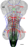
Overall neurovascular pattern in adult goldfish. Reconstruction of adult goldfish hindbrain retrogradely labeled from spinal cord with dextran‐conjugated biotin. Schematic overlay of blue rhombomeric (1–8) boundaries, green motor nuclei and red central arterial stems and branches. CA, central arteries of the hindbrain; III‐X, cranial motor nuclei; MB, midbrain; Mes, mesencephalic arteries; OLE, octavolateral efferent nucleus; OS, occipito‐spinal motor column; r, rhombomere; SC, spinal cord; VL, vagal lobe. Scale bar: 1 mm.

Similar articles

Cited by

References

    1. Allen W (1905) . The blood-vascular system of the Loricati, the mail-cheeked fishes. Proc Washington Acad Sci 7,27‐157
    1. Allis E (1912) The pseudobranchial and carotid arteries in Esox, Salmo and Gadus, together with a description of the arteries of the adult Amia. Anat Anz 41, 113–141.
    1. Anadón R, Molist P, Rodríguez‐Moldes I, et al. (2000) Distribution of choline acetyltransferase immunoreactivity in the brain of an elasmobranch, the lesser spotted dogfish (Scyliorhinus canicula). J Comp Neurol 420, 139–170. - PubMed
    1. Bass AH, Gilland E, Baker R (2008) Evolutionary origins for social vocalization in a vertebrate hindbrain‐spinal compartment. Science 321, 417–421. - PMC - PubMed
    1. Brantley RK, Bass AH (1988) Cholinergic neurons in the brain of a teleost fish (Porichthys notatus) located with an antibody to choline acetyltransferase. J Comp Neurol 275, 87–105. - PubMed

Publication types

MeSH terms

LinkOut - more resources