Review
. 2020 Jun;32(18):1330-1347.
doi: 10.1089/ars.2019.7803. Epub 2019 Jul 18.

Metabolic Responses to Reductive Stress

Affiliations
Review

Metabolic Responses to Reductive Stress

Wusheng Xiao et al. Antioxid Redox Signal. 2020 Jun.

Abstract

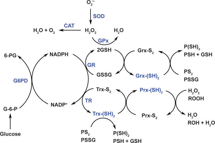
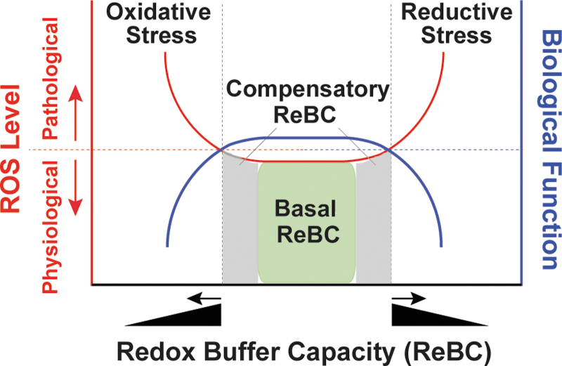
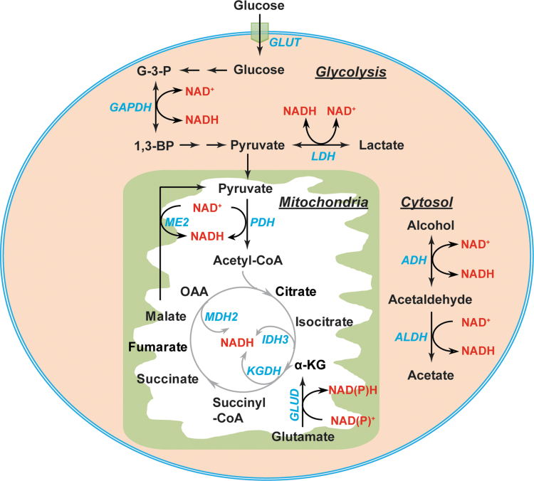
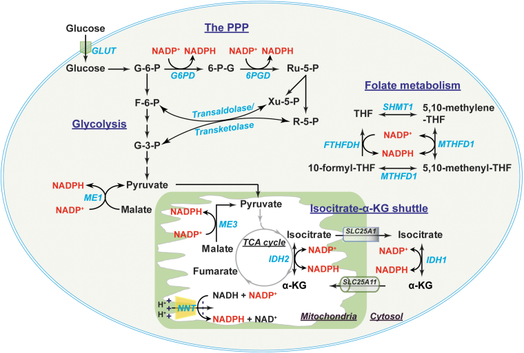
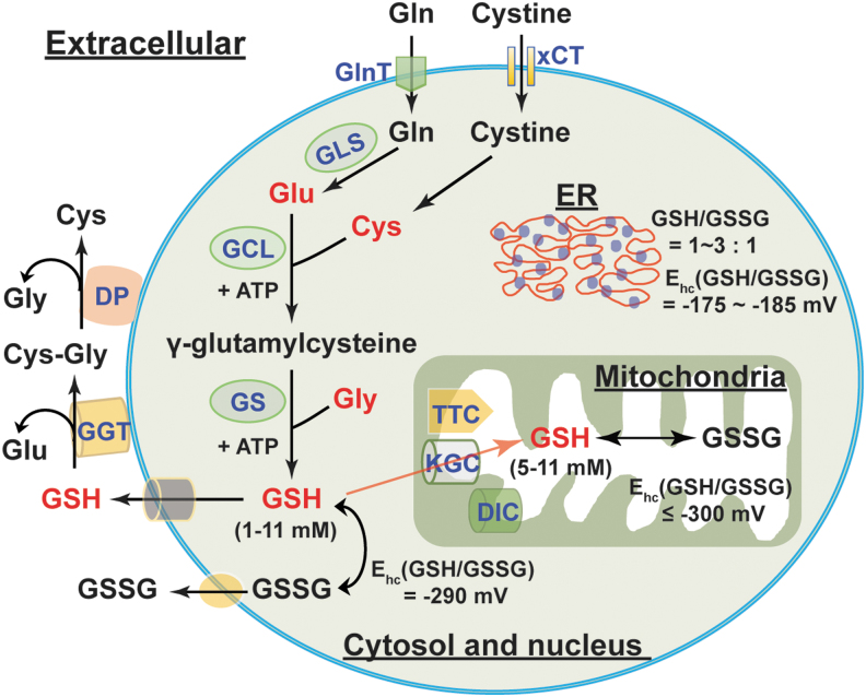
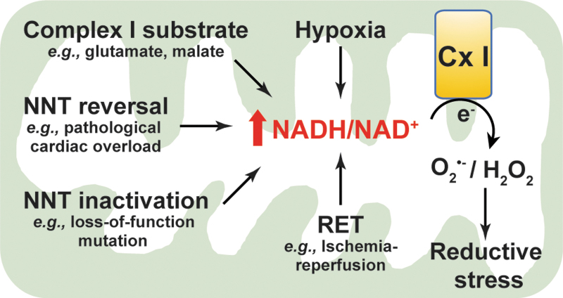
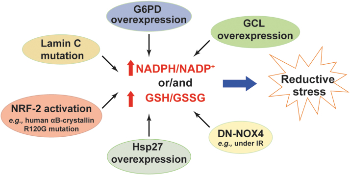
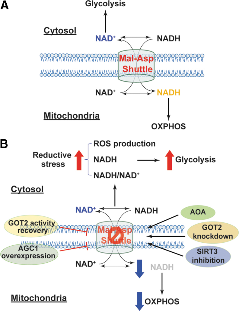
Significance: Reducing equivalents (NAD(P)H and glutathione [GSH]) are essential for maintaining cellular redox homeostasis and for modulating cellular metabolism. Reductive stress induced by excessive levels of reduced NAD+ (NADH), reduced NADP+ (NADPH), and GSH is as harmful as oxidative stress and is implicated in many pathological processes. Recent Advances: Reductive stress broadens our view of the importance of cellular redox homeostasis and the influences of an imbalanced redox niche on biological functions, including cell metabolism. Critical Issues: The distribution of cellular NAD(H), NADP(H), and GSH/GSH disulfide is highly compartmentalized. Understanding how cells coordinate different pools of redox couples under unstressed and stressed conditions is critical for a comprehensive view of redox homeostasis and stress. It is also critical to explore the underlying mechanisms of reductive stress and its biological consequences, including effects on energy metabolism. Future Directions: Future studies are needed to investigate how reductive stress affects cell metabolism and how cells adapt their metabolism to reductive stress. Whether or not NADH shuttles and mitochondrial nicotinamide nucleotide transhydrogenase enzyme can regulate hypoxia-induced reductive stress is also a worthy pursuit. Developing strategies (e.g., antireductant approaches) to counteract reductive stress and its related adverse biological consequences also requires extensive future efforts.

Keywords: GSH/GSSG; NAD(H); NADP(H); cellular metabolism; redox; reductive stress.


Figures

FIG. 1.
FIG. 1.
Cellular redox network. Cellular redox homeostasis is maintained by a delicate balance between pro-oxidant and antioxidant systems. Cellular pro-oxidants primarily include O2•− and H2O2, which are generated by enzymes or/and mitochondrial respiration. SODs convert O2•− to H2O2, which is further neutralized to water by catalase, GPxs, and Prxs. Catalase has very low affinity for H2O2 (but with a high turnover and high catalytic efficiency) and can reduce H2O2 to water when its levels reach the millimolar range. GPxs require two molecules of reduced GSH for H2O2 reduction and GSH is oxidized to GSSG in this reaction. GSH can also be used by glutaredoxins [Grx-(SH)2] to reduce protein intra/inter-disulfide (PS2 or PSSG) into reduced protein cysteinyl residues (P[SH]2 or PSH + GSH). The recycling of GSSG to GSH is catalyzed by GR, which utilizes NADPH as an electron donor. Prxs extract electrons from reduced thioredoxins [Trx-(SH)2] to reduce H2O2 or organic hydroperoxides (ROOH) to H2O or/and alcohols (ROH), respectively; and Trx-(SH)2 is simultaneously oxidized into Trx-S2. Like GSH, Trx-(SH)2 can also reduce protein disulfides into protein cysteinyls. The recycling of Trx-S2 is catalyzed by TRs using NADPH as an electron donor. NADPH is generated by G6PD in the PPP of glucose metabolism. Therefore, the cellular redox state and cell metabolism are closely linked through NADPH. Adapted from references (7, 8) with permission. 6-PG, 6-phosphogluconate; G-6-P, glucose-6-phosphate; G6PD, glucose-6-phosphate dehydrogenase; GPxs, glutathione peroxidases; GR, glutathione reductase; Grx, glutaredoxin; GSH, glutathione; GSSG, GSH disulfide; H2O2, hydrogen peroxide; NADPH, reduced NADP+; O2•−, superoxide anion; PPP, pentose phosphate pathway; Prxs, peroxiredoxins; PSSG, protein-glutathione disulfide; SODs, superoxide dismutases; TRs, thioredoxin reductases; Trx, thioredoxin.
FIG. 2.
FIG. 2.
Redox stress. The NADH/NAD+, NADPH/NADP+, and GSH/GSSG redox couples are the major cellular redox buffers. Under unstressed conditions, these three redox couples have adequate capacity to maintain redox homeostasis, termed basal ReBC (X-axis; green box under red line). When cellular ROS levels increase (Left Y-axis; red line), these redox buffers are also able to respond by elevating basal ReBC to a certain level, termed the compensatory ReBC (X-axis; gray area under red line). Under both circumstances, cellular ROS levels are maintained at physiological levels to ensure normal biological function, such as signaling molecule adequacy (Right Y-axis; blue line). However, when this compensatory response continues beyond a certain threshold at which the maximal ReBC is exceeded by cellular reducing of ROS levels (X-axis, black triangle), reductive stress can occur (red line). By contrast, oxidative stress occurs when cellular ReBC decreases (X-axis, black triangle), or/and cellular oxidation of ROS production is overwhelming. Both reductive stress and oxidative stress (collectively named as redox stress) can promote ROS production leading to oxidative damages to macromolecules and perturbations of cellular functions. NAD+, nicotinamide adenine dinucleotide; NADH, reduced NAD+; NADP+, phosphorylated NAD+; ReBC, redox buffer capacity; ROS, reactive oxygen species.
FIG. 3.
FIG. 3.
Metabolic sources of NAD(H). In the cytosol, the interconversion of NAD+ and NADH is mediated by two enzymes GAPDH and LDH and by the alcohol metabolism enzymes ADH and ALDH. In the mitochondrial matrix, PDH, ME2, GLUD, and TCA cycle enzymes (IDH3, KGDH, and MDH2) contribute to NAD(H) production. 1,3-BPG, 1,3-bisphosphoglycerate; α-KG, α-ketoglutarate; ADH, alcohol dehydrogenase; ALDH, aldehyde dehydrogenase; G-3-P, glyceraldehyde-3-phosphate; GAPDH, glyceraldehyde-3-phosphate dehydrogenase; GLUD, glutamate dehydrogenase; GLUT, glucose transporter; IDH, isocitrate dehydrogenase; KGDH, α-ketoglutarate dehydrogenase; LDH, lactate dehydrogenase; MDH2, malate dehydrogenase 2; ME, malic enzyme; OAA, oxaloacetate; PDH, pyruvate dehydrogenase; TCA, tricarboxylic acid.
FIG. 4.
FIG. 4.
Metabolic sources of NADP(H) and the NADPH shuttle. In the cytosol, NADPH is primarily produced by G6PD and 6PGD in the PPP, where the products R-5-P and Xu-5-P can be diverted into glycolysis by transaldolase and transketolase. ME1 and IDH1 also contribute to cytosolic NADP(H) production. In addition, two enzymes in folate metabolism, MTHFD1 and FTHFDH, are also the sources of cytosolic NADP(H). Mitochondrial NADP(H) is generated by NADP+-dependent IDH2, NNT, and ME3. The cytosolic and mitochondrial NADP(H) pools are exchanged through the isocitrate-α-KG shuttle, where cytosolic IDH1 and mitochondrial IDH2 catalyze the interconversion of isocitrate and α-KG in conjunction with the interconversion of NADP+ and NADPH. The citrate carrier protein (encoded by SLC25A1 gene) and the α-KG/malate antiporter (encoded by SLC25A11 gene) mediate the transport of isocitrate and α-KG between the cytosol and mitochondria, respectively. 6PGD, 6-phosphogluconate dehydrogenase; FTHFDH, 10-formyl-tetrahydrofolate dehydrogenase; MTHFD, methylene-tetrahydrofolate dehydrogenase; NNT, nicotinamide nucleotide transhydrogenase; R-5-P, ribose-5-phosphate; Ru-5-P, ribulose-5-phosphate; SCL25A1, solute carrier family 25 member 1; SCL25A11, solute carrier family 25 member 11; SHMT, serine hydroxymethyltransferase; THF, tetrahydrofolate; Xu-5-P, xylulose-5-phosphate.
FIG. 5.
FIG. 5.
The biosynthesis and cellular distribution of GSH. Glu, Cys, and Gly are three precursor amino acids of GSH. GSH synthesis requires two successive steps catalyzed by two ATP-dependent enzymes: GCL (the rate-limiting enzyme) and GS. Glu can be replenished by endogenous and exogenous Gln via GLS-mediated glutaminolysis. Cys can be generated by reduction of endogenous and exogenous cystine. GSH levels in the cytosol and nucleus are within the 1–11 mM range with a half-cell reduction potential (Ehc) of −290 mV. By contrast, the ER has a more oxidizing environment with GSH/GSSG of 1–3:1 and an Ehc of −175 to −185 mV. Cytosolic GSH can be transported into mitochondria through three potential transporters, KGC, DIC, and TTC. Mitochondrial GSH levels and the Ehc are comparable with that of cytosolic and nuclear pools. In addition, GSH and GSSG can also be secreted into the extracellular compartment; however, their levels are very low comparatively (within micromolar range). Extracellular GSH can be degraded by the cell surface enzyme GGT to form Glu and Cys-Gly, further metabolized into free Cys and Gly by DP. The resulting amino acids can be reused by cells for intracellular GSH synthesis. Cys, cysteine; Cys-Gly, cysteinyl-glycine; DIC, dicarboxylate carrier; DP, dipeptidases; ER, endoplasmic reticulum; GCL, γ-glutamylcysteine ligase; GGT, γ-glutamyl transpeptidase; GLS, glutaminase; Glu, glutamate; Gly, glycine; GS, GSH synthetase; KGC, α-KG carrier; TTC, tricarboxylate carrier; xCT, cystine transporter.
FIG. 6.
FIG. 6.
Excess in NADH levels induces reductive stress. Under stressed states, such as exogenous addition of Cx I substrates, hypoxia, NNT reversal, NNT inactivation, and RET, mitochondrial NADH/NAD+ increases leading to one-electron reduction of oxygen to O2•− or/and two-electron reduction of oxygen into H2O2 at respiratory Cx I. Extensive reductive ROS levels result in reductive stress, which is detrimental to cellular function. Cx I, complex I; RET, reverse electron transfer.
FIG. 7.
FIG. 7.
Excess in NADPH or/and GSH levels triggers reductive stress. Increases in NADPH/NADP+ or/and GSH/GSSG due to increases in their production (e.g., G6PD overexpression, NRF-2 activation, Hsp27 overexpression, GCL overexpression, and lamin C mutation) or decreases in their consumption (e.g., overexpression of DN-NOX4) lead to reductive stress. DN-NOX4, dominant negative NADPH oxidase 4; Hsp27, heat shock protein 27; IR, ischemia/reperfusion; NRF-2, nuclear factor (erythroid-derived 2)-like 2.
FIG. 8.
FIG. 8.
Inhibition of the malate/aspartate NADH shuttle induces reductive stress and metabolic reprogramming. (A) The malate/aspartate shuttle exchanges cytosolic NAD(H) with mitochondrial NAD(H) to maintain high cytosolic NAD+ levels, which are required for glycolysis, and high mitochondrial NADH levels, which provide electrons for mitochondrial OXPHOS. (B) Inhibition of the malate/aspartate shuttle by silencing GOT2, suppressing SIRT3 activity, or using the chemical inhibitor AOA leads to accumulation of cytosolic NADH and increases in cytosolic NADH/NAD+ and reductive ROS generation, indicative of reductive stress. NADH-induced reductive stress shifts cell metabolism from mitochondrial respiration to glycolysis. Enhancement of this shuttle activity by overexpression of AGC1 or recovery of GOT2 activity significantly increases mitochondrial NADH levels and enhances mitochondrial respiration. AGC1, aspartate-glutamate carrier 1; AOA, aminooxyacetic acid; GOT2, glutamate-oxaloacetate transaminase; Mal-Asp shuttle, malate/aspartate shuttle; OXPHOS, oxidative phosphorylation; SIRT3, sirtuin deacetylase family member 3.

References

    1. Alderson NL, Wang Y, Blatnik M, Frizzell N, Walla MD, Lyons TJ, Alt N, Carson JA, Nagai R, Thorpe SR, and Baynes JW. S-(2-Succinyl)cysteine: a novel chemical modification of tissue proteins by a Krebs cycle intermediate. Arch Biochem Biophys 450: 1–8, 2006 - PubMed
    1. Aquilano K, Baldelli S, and Ciriolo MR. Glutathione: new roles in redox signaling for an old antioxidant. Front Pharmacol 5: 196, 2014 - PMC - PubMed
    1. Barron JT, Gu L, and Parrillo JE. Malate-aspartate shuttle, cytoplasmic NADH redox potential, and energetics in vascular smooth muscle. J Mol Cell Cardiol 30: 1571–1579, 1998 - PubMed
    1. Barron JT, Gu L, and Parrillo JE. NADH/NAD redox state of cytoplasmic glycolytic compartments in vascular smooth muscle. Am J Physiol Heart Circ Physiol 279: H2872–H2878, 2000 - PubMed
    1. Bedard K and Krause KH. The NOX family of ROS-generating NADPH oxidases: physiology and pathophysiology. Physiol Rev 87: 245–313, 2007 - PubMed

Publication types