Review
. 2019 Jun 20;15(6):e1006648.
doi: 10.1371/journal.pcbi.1006648. eCollection 2019 Jun.

Protein ensembles link genotype to phenotype

Affiliations
Review

Protein ensembles link genotype to phenotype

Ruth Nussinov et al. PLoS Comput Biol. .

Abstract

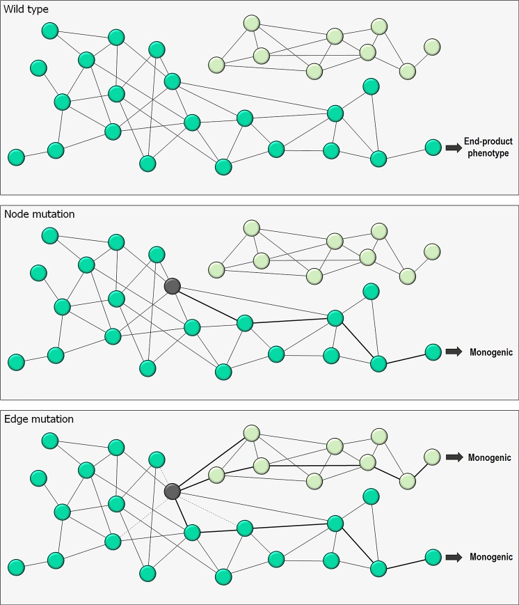
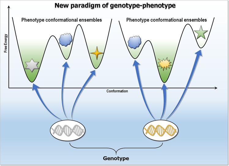
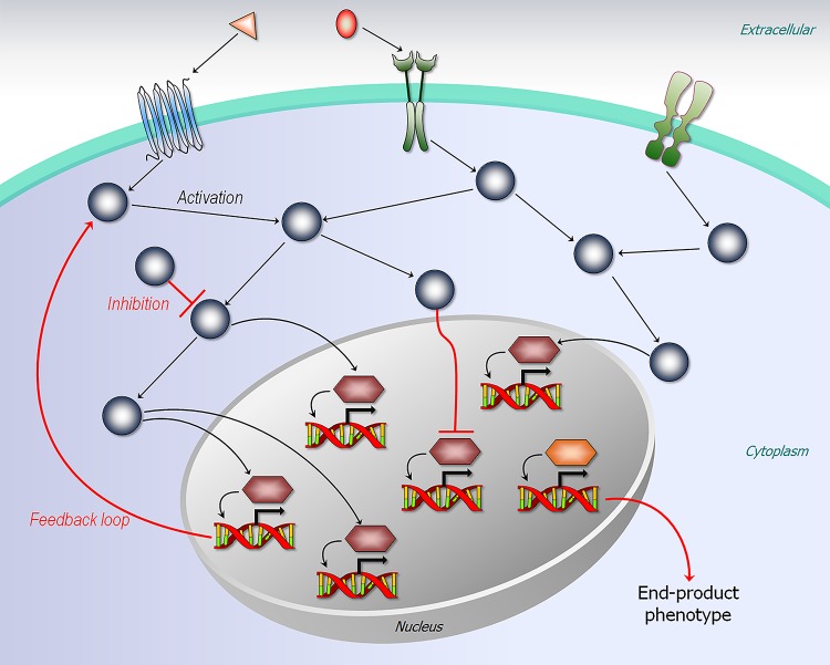
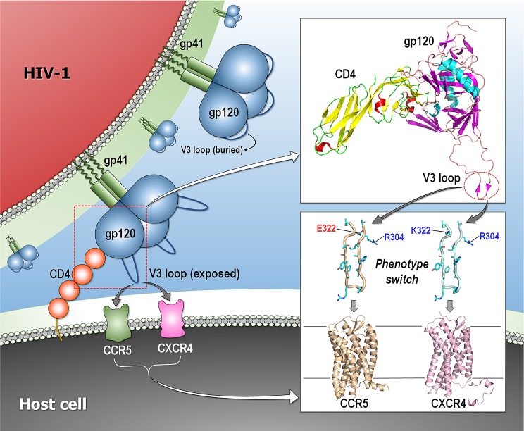
Classically, phenotype is what is observed, and genotype is the genetic makeup. Statistical studies aim to project phenotypic likelihoods of genotypic patterns. The traditional genotype-to-phenotype theory embraces the view that the encoded protein shape together with gene expression level largely determines the resulting phenotypic trait. Here, we point out that the molecular biology revolution at the turn of the century explained that the gene encodes not one but ensembles of conformations, which in turn spell all possible gene-associated phenotypes. The significance of a dynamic ensemble view is in understanding the linkage between genetic change and the gained observable physical or biochemical characteristics. Thus, despite the transformative shift in our understanding of the basis of protein structure and function, the literature still commonly relates to the classical genotype-phenotype paradigm. This is important because an ensemble view clarifies how even seemingly small genetic alterations can lead to pleiotropic traits in adaptive evolution and in disease, why cellular pathways can be modified in monogenic and polygenic traits, and how the environment may tweak protein function.


Conflict of interest statement

The authors have declared that no competing interests exist.

Figures

Fig 1
Fig 1. Classical view of genotype–phenotype.
In this view, a protein or the gene product is considered to have one shape with a single function. Monogenic traits are expressed by single genes, whereas polygenic traits are affected by multiple genes. Seemingly unrelated phenotypic traits are pleiotropy that can be expressed by a single gene.
Fig 2
Fig 2. Network perturbations of genotype–phenotype.
Mutations can shift phenotype traits generated from wild-type trait (upper panel), affecting solely the protein (a “node”) or protein–protein interaction (an “edge”). The former and latter are node and edge mutations, respectively. The node mutation (middle panel) generates a monogenetic trait, whereas the edge mutation (bottom panel) creates or breaks the protein–protein interactions yielding monogenetic or polygenetic traits. An example shown for edge trait is polygenetic.
Fig 3
Fig 3. New paradigm of genotype–phenotype.
In this view, genotype encodes a distinct conformational ensemble in all states. Populations determine the specific phenotype traits that link to genotype. Mutations shift the equilibrium of preexisting conformational ensembles altering phenotypes.
Fig 4
Fig 4. Cellular network.
The network controls the transcriptional regulation for gene. Edge mutations can alter the cellular network, expressing the end-product phenotype.
Fig 5
Fig 5. Phenotype switch of HIV-1 entry.
Schematic diagram representing the initial process of the HIV-1 entry that led to fusion between the viral and the host cell membranes. The gp120 trimer undergoes a conformational change upon binding to cellular receptor CD4, exposing the variable loop V3. The V3 loop binds to the coreceptor (CCR5 and CXCR4), triggering the entry process. The wild-type V3 loop with positively and negatively charged residues at positions 304 and 322, respectively, recognizes CCR5. A phonotype switch by a mutation from a negatively to a positively charged residue at position 322 in the V3 loop alters coreceptor recognition to CXCR4. Modeled structures are the crystal structures of gp120 (PDB code: 24BC), CCR5 (PDB code: 4MBS), and CXCR4 (PDB code: 3ODU) and the NMR structure of V3 loop (PDB code: 2ESX). CCR5, C-C chemokine receptor type 5; CXCR4, C-X-C chemokine receptor type 4; PDB, Protein Data Bank.

Similar articles

Cited by

References

    1. Lewontin RC (1985) Population genetics. Annu Rev Genet 19:81–102. 10.1146/annurev.ge.19.120185.000501 - DOI - PubMed
    1. Nussinov R, Wolynes PG (2014) A second molecular biology revolution? The energy landscapes of biomolecular function. Phys Chem Chem Phys 16(14):6321–6322. 10.1039/c4cp90027h - DOI - PubMed
    1. Frauenfelder H, Sligar SG, Wolynes PG (1991) The energy landscapes and motions of proteins. Science 254(5038):1598–1603. https://10.1126/science.1749933 - DOI - PubMed
    1. Tsai CJ, Kumar S, Ma B, Nussinov R (1999) Folding funnels, binding funnels, and protein function. Protein Sci 8(6):1181–1190. 10.1110/ps.8.6.1181 - DOI - PMC - PubMed
    1. Ma B, Kumar S, Tsai CJ, Nussinov R (1999) Folding funnels and binding mechanisms. Protein Eng 12(9):713–720. https://10.1093/protein/12.9.713 - DOI - PubMed

Publication types