

Green synthesis of silver nanoparticles via Cynara scolymus leaf extracts: The characterization, anticancer potential with photodynamic therapy in MCF7 cells
- PMID: 31220110
- PMCID: PMC6586393
- DOI: 10.1371/journal.pone.0216496
Green synthesis of silver nanoparticles via Cynara scolymus leaf extracts: The characterization, anticancer potential with photodynamic therapy in MCF7 cells
Abstract
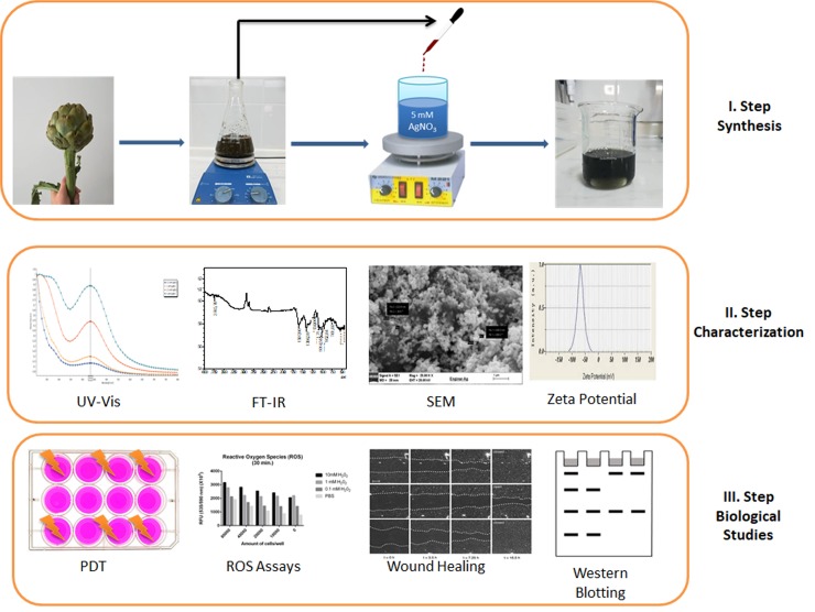
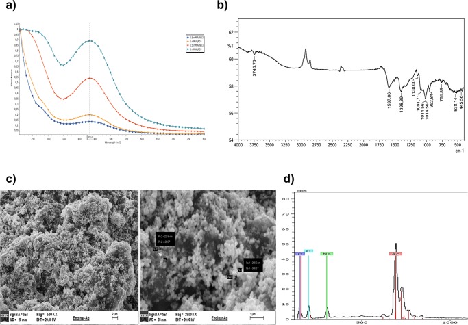
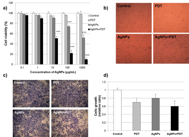
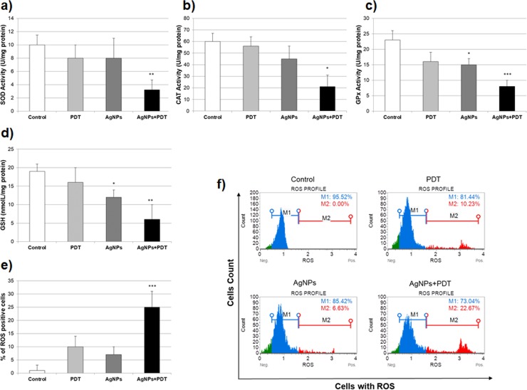
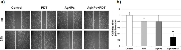
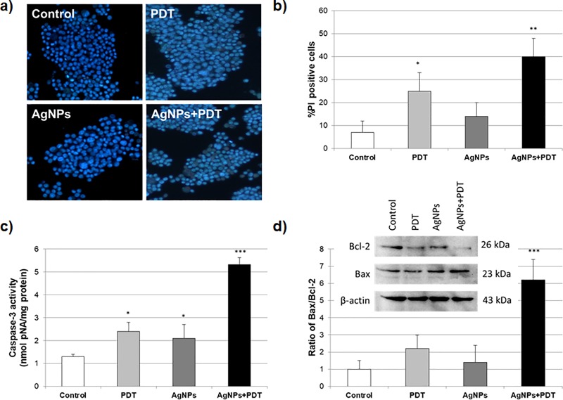
In this study, we report on the synthesis of silver nanoparticles (AgNPs) from the leaf extracts of Cynara scolymus (Artichoke) using microwave irradiation and the evaluation of its anti-cancer potential with photodynamic therapy (PDT). Silver nanoparticles formation was characterized by scanning electron microscopy with energy dispersive x-ray spectroscopy and Fourier transform infrared (FTIR) spectroscopy. Silver nanoparticles formation was also investigated the surface charge, particle size and distribution using zetasizer analysis. The cytotoxic effect of AgNPs and/or PDT was studied by MTT assay and migration by the scratch assay. The apoptotic inducing ability of the AgNPs and/or PDT was investigated by intracellular ROS analysis, antioxidant enzyme levels (SOD, CAT, GPx and GSH), Hoechst staining and Bax/Bcl-2 analysis using western blotting. The mean particle size of produced AgNPs was found 98.47±2.04 nm with low polydispersity (0.301±0.033). Zeta potential values of AgNPs show -32.3± 0.8 mV. These results clearly indicate the successful formation of AgNPs for cellular uptake. Mitochondrial damage and intracellular ROS production were observed upon treatment with AgNPs (10μg/mL) and PDT (0.5 mJ/cm2) showed significant reducing cell migration, expression of Bax and suppression of Bcl-2. Significantly, biosynthesized AgNPs showed a broad-spectrum anti-cancer activity with PDT therapy and therefore represent promoting ROS generation by modulating mitochondrial apoptosis induction in MCF7 breast cancer cells.
Conflict of interest statement
The authors have declared that no competing interests exist.
Figures
Similar articles
-
Insight into the molecular mechanism, cytotoxic, and anticancer activities of phyto-reduced silver nanoparticles in MCF-7 breast cancer cell lines.Microsc Res Tech. 2024 Jul;87(7):1627-1639. doi: 10.1002/jemt.24540. Epub 2024 Mar 7. Microsc Res Tech. 2024. PMID: 38450823
-
Silver nanoparticles biosynthesised using Centella asiatica leaf extract: apoptosis induction in MCF-7 breast cancer cell line.IET Nanobiotechnol. 2018 Oct;12(7):994-1002. doi: 10.1049/iet-nbt.2018.5069. IET Nanobiotechnol. 2018. PMID: 30247143 Free PMC article.
-
Eco-friendly green synthesis of silver nanoparticles and their potential applications as antioxidant and anticancer agents.Drug Dev Ind Pharm. 2019 Oct;45(10):1682-1694. doi: 10.1080/03639045.2019.1656224. Epub 2019 Sep 2. Drug Dev Ind Pharm. 2019. PMID: 31407925
-
Bioinspired and Green Synthesis of Silver Nanoparticles for Medical Applications: A Green Perspective.Appl Biochem Biotechnol. 2024 Jun;196(6):3636-3669. doi: 10.1007/s12010-023-04719-z. Epub 2023 Sep 5. Appl Biochem Biotechnol. 2024. PMID: 37668757 Free PMC article. Review.
-
Pharmaceutical Aspects of Green Synthesized Silver Nanoparticles: A Boon to Cancer Treatment.Anticancer Agents Med Chem. 2021;21(12):1490-1509. doi: 10.2174/1871520620666200918111024. Anticancer Agents Med Chem. 2021. PMID: 32951580 Review.
Cited by
-
Biologically Synthesized Silver Nanoparticles and Their Diverse Applications.Nanomaterials (Basel). 2022 Sep 9;12(18):3126. doi: 10.3390/nano12183126. Nanomaterials (Basel). 2022. PMID: 36144915 Free PMC article. Review.
-
In vitro hepatotoxicity evaluation of methotrexate-loaded niosome formulation: fabrication, characterization and cell culture studies.Turk J Med Sci. 2023 Aug;53(4):872-882. doi: 10.55730/1300-0144.5651. Epub 2023 Aug 18. Turk J Med Sci. 2023. PMID: 38031943 Free PMC article.
-
The Potential of Antibody Technology and Silver Nanoparticles for Enhancing Photodynamic Therapy for Melanoma.Biomedicines. 2022 Sep 1;10(9):2158. doi: 10.3390/biomedicines10092158. Biomedicines. 2022. PMID: 36140259 Free PMC article. Review.
-
Synthesis of halloysite nanotubes decorated with green silver nanoparticles to investigate cytotoxicity, lipid peroxidation and induction of apoptosis in acute leukemia cells.Sci Rep. 2023 Oct 11;13(1):17182. doi: 10.1038/s41598-023-43978-y. Sci Rep. 2023. PMID: 37821481 Free PMC article.
-
Novel biogenic silver nanoconjugates of Abrus precatorius seed extracts and their antiproliferative and antiangiogenic efficacies.Sci Rep. 2023 Aug 19;13(1):13514. doi: 10.1038/s41598-023-40079-8. Sci Rep. 2023. PMID: 37598190 Free PMC article.
References
-
- Lopes CRB, Courrol LC. Green synthesis of silver nanoparticles with extract of Mimusops coriacea and light. Journal of Luminescence. 2018;199:183–7. 10.1016/j.jlumin.2018.03.030. - DOI
-
- Saratale RG, Karuppusamy I, Saratale GD, Pugazhendhi A, Kumar G, Park Y, et al. A comprehensive review on green nanomaterials using biological systems: Recent perception and their future applications. Colloids and Surfaces B: Biointerfaces. 2018;170:20–35. 10.1016/j.colsurfb.2018.05.045 - DOI - PubMed
Publication types
MeSH terms
Substances
LinkOut - more resources
Full Text Sources
Research Materials
Miscellaneous
