

The Oxysterol Synthesising Enzyme CH25H Contributes to the Development of Intestinal Fibrosis
- PMID: 31220227
- PMCID: PMC6751338
- DOI: 10.1093/ecco-jcc/jjz039
The Oxysterol Synthesising Enzyme CH25H Contributes to the Development of Intestinal Fibrosis
Abstract
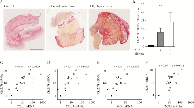
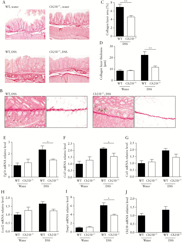
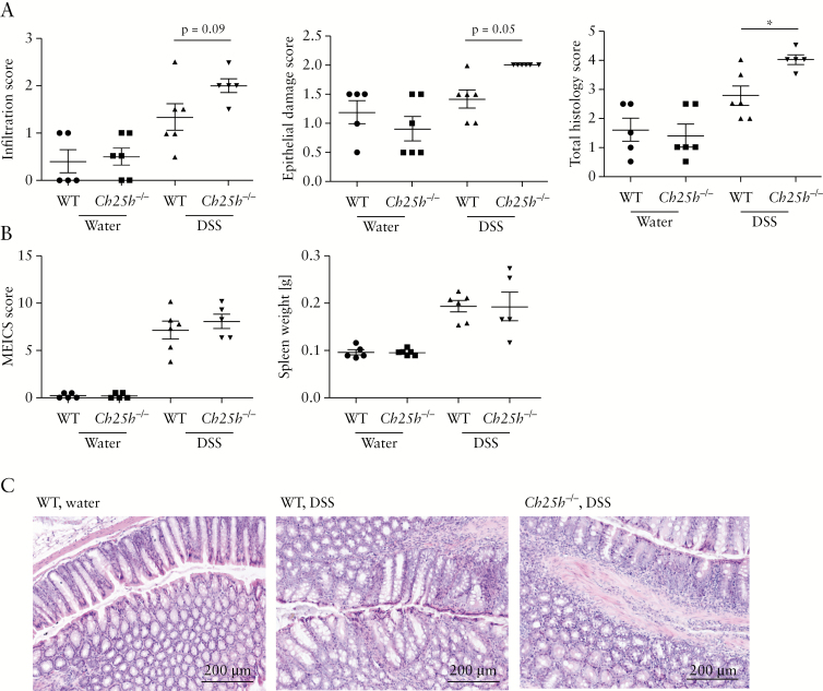
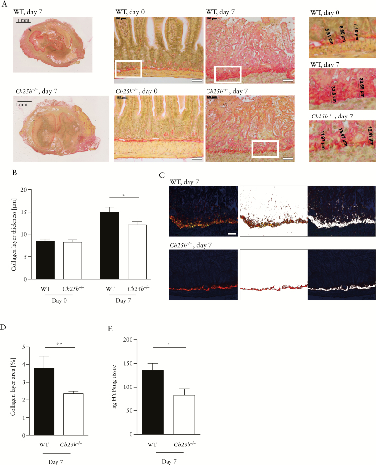
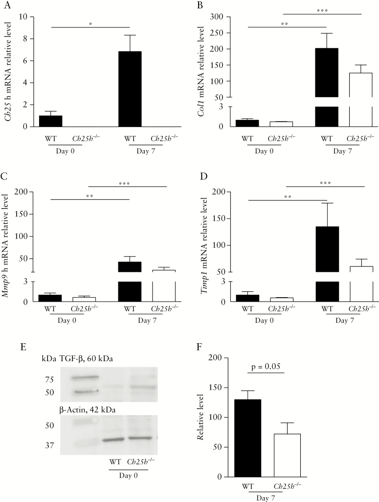
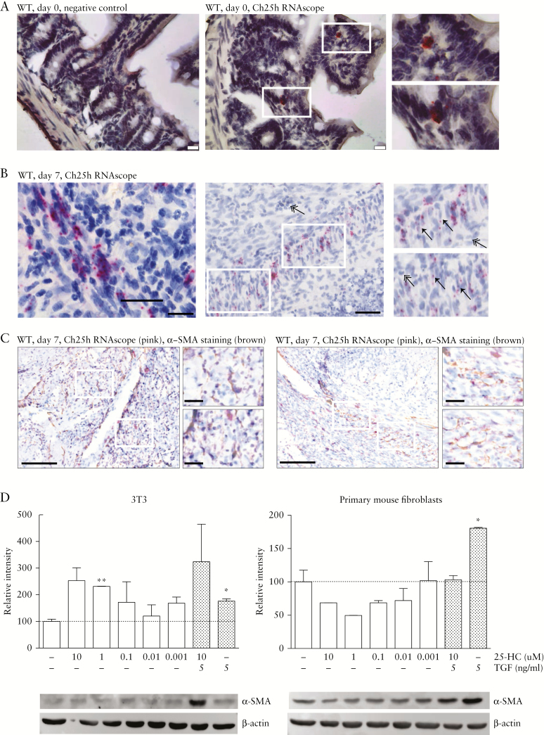
Intestinal fibrosis and stenosis are common complications of Crohn's disease [CD], frequently requiring surgery. Anti-inflammatory strategies can only partially prevent fibrosis; hence, anti-fibrotic therapies remain an unmet clinical need. Oxysterols are oxidised cholesterol derivatives with important roles in various biological processes. The enzyme cholesterol 25-hydroxylase [CH25H] converts cholesterol to 25-hydroxycholesterol [25-HC], which modulates immune responses and oxidative stress. In human intestinal samples from CD patients, we found a strong correlation of CH25H mRNA expression with the expression of fibrosis markers. We demonstrate reduced intestinal fibrosis in mice deficient for the CH25H enzyme, using the sodium dextran sulphate [DSS]-induced chronic colitis model. Additionally, using a heterotopic transplantation model of intestinal fibrosis, we demonstrate reduced collagen deposition and lower concentrations of hydroxyproline in CH25H knockouts. In the heterotopic transplant model, CH25H was expressed in fibroblasts. Taken together, our findings indicate an involvement of oxysterol synthesis in the pathogenesis of intestinal fibrosis.
Keywords: Fibrogenesis; cholesterol 25 hydroxylase [CH25H]; graft; intestinal fibrosis; mouse model; oxysterols; transplantation.
© European Crohn’s and Colitis Organisation (ECCO) 2019.
Figures
Similar articles
-
Research progress on the mechanism of cholesterol-25-hydroxylase in intestinal immunity.Front Immunol. 2023 Aug 31;14:1241262. doi: 10.3389/fimmu.2023.1241262. eCollection 2023. Front Immunol. 2023. PMID: 37720208 Free PMC article. Review.
-
Cholesterol 25-hydroxylase protects against experimental colitis in mice by modulating epithelial gut barrier function.Sci Rep. 2020 Aug 28;10(1):14246. doi: 10.1038/s41598-020-71198-1. Sci Rep. 2020. PMID: 32859970 Free PMC article.
-
Cholesterol 25-hydroxylase (CH25H) as a promoter of adipose tissue inflammation in obesity and diabetes.Mol Metab. 2020 Sep;39:100983. doi: 10.1016/j.molmet.2020.100983. Epub 2020 Mar 27. Mol Metab. 2020. PMID: 32229247 Free PMC article.
-
Severity of local inflammation does not impact development of fibrosis in mouse models of intestinal fibrosis.Sci Rep. 2018 Oct 12;8(1):15182. doi: 10.1038/s41598-018-33452-5. Sci Rep. 2018. PMID: 30315190 Free PMC article.
-
Fibrogenesis. IV. Fibrosis and inflammatory bowel disease: cellular mediators and animal models.Am J Physiol Gastrointest Liver Physiol. 2000 Oct;279(4):G653-9. doi: 10.1152/ajpgi.2000.279.4.G653. Am J Physiol Gastrointest Liver Physiol. 2000. PMID: 11005750 Review.
Cited by
-
The Protective Effect of Dabigatran and Rivaroxaban on DNA Oxidative Changes in a Model of Vascular Endothelial Damage with Oxidized Cholesterol.Int J Mol Sci. 2020 Mar 13;21(6):1953. doi: 10.3390/ijms21061953. Int J Mol Sci. 2020. PMID: 32182973 Free PMC article.
-
Genomics and metabolomics of early-stage thioacetamide-induced liver injury: An interspecies study between guinea pig and rat.Toxicol Appl Pharmacol. 2021 Nov 1;430:115713. doi: 10.1016/j.taap.2021.115713. Epub 2021 Sep 4. Toxicol Appl Pharmacol. 2021. PMID: 34492290 Free PMC article.
-
Current Approach to Risk Factors and Biomarkers of Intestinal Fibrosis in Inflammatory Bowel Disease.Medicina (Kaunas). 2024 Feb 10;60(2):305. doi: 10.3390/medicina60020305. Medicina (Kaunas). 2024. PMID: 38399592 Free PMC article. Review.
-
Effects of Oxysterols on Immune Cells and Related Diseases.Cells. 2022 Apr 7;11(8):1251. doi: 10.3390/cells11081251. Cells. 2022. PMID: 35455931 Free PMC article. Review.
-
Research progress on the mechanism of cholesterol-25-hydroxylase in intestinal immunity.Front Immunol. 2023 Aug 31;14:1241262. doi: 10.3389/fimmu.2023.1241262. eCollection 2023. Front Immunol. 2023. PMID: 37720208 Free PMC article. Review.
References
-
- Bernstein CN, Loftus EV Jr, Ng SC, Lakatos PL, Moum B; Epidemiology and Natural History Task Force of the International Organization for the Study of Inflammatory Bowel Disease [IOIBD] Hospitalisations and surgery in Crohn’s disease. Gut 2012;61:622–9. - PubMed
-
- Bemelman WA, Allez M. The surgical intervention: earlier or never? Best Pract Res Clin Gastroenterol 2014;28:497–503. - PubMed
-
- Gionchetti P, Dignass A, Danese S, et al. ; ECCO Third European evidence-based consensus on the diagnosis and management of Crohn’s disease 2016. Part 2: surgical management and special situations. J Crohns Colitis 2017;11:135–49. - PubMed
-
- Gordon IO, Agrawal N, Goldblum JR, Fiocchi C, Rieder F. Fibrosis in ulcerative colitis: mechanisms, features, and consequences of a neglected problem. Inflamm Bowel Dis 2014;20:2198–206. - PubMed
MeSH terms
Substances
LinkOut - more resources
Full Text Sources
