. 2020 Feb;182(2):468-476.
doi: 10.1111/bjd.18239. Epub 2019 Oct 27.

Basal cell carcinoma characterization using fusion ex vivo confocal microscopy: a promising change in conventional skin histopathology

Affiliations

Basal cell carcinoma characterization using fusion ex vivo confocal microscopy: a promising change in conventional skin histopathology

J Pérez-Anker et al. Br J Dermatol. 2020 Feb.

Abstract

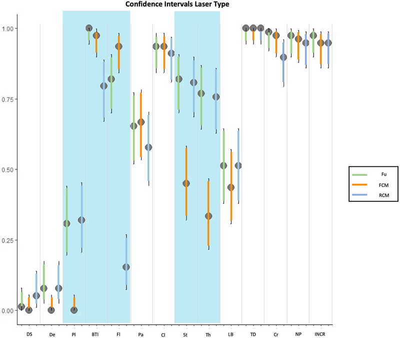
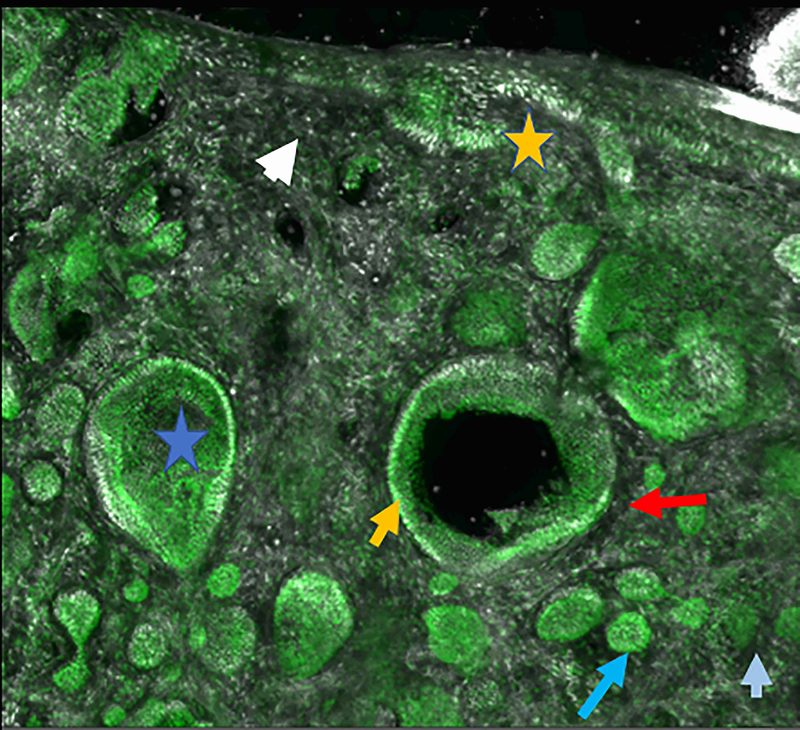
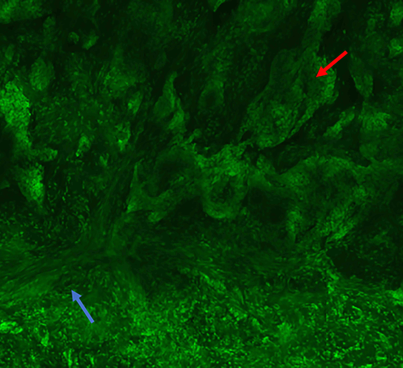
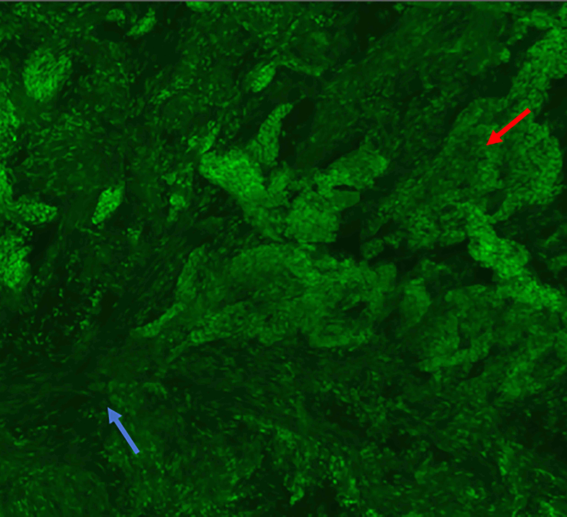
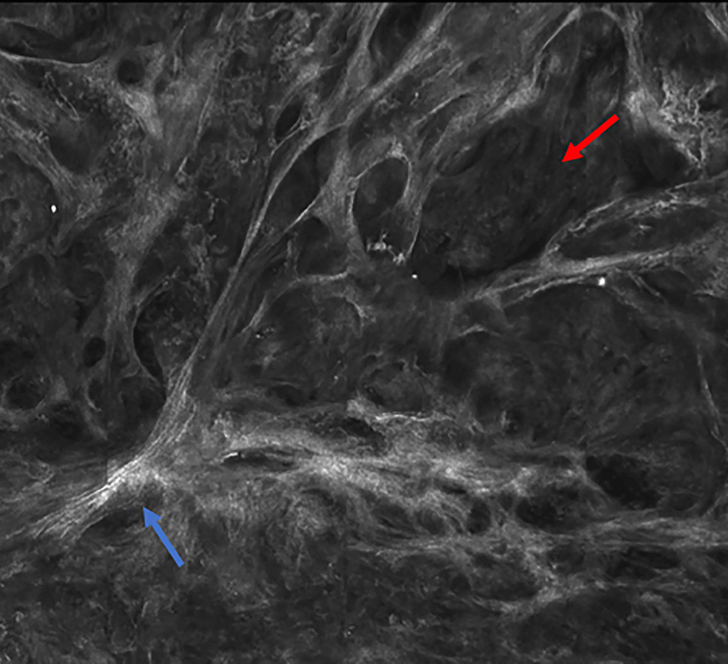
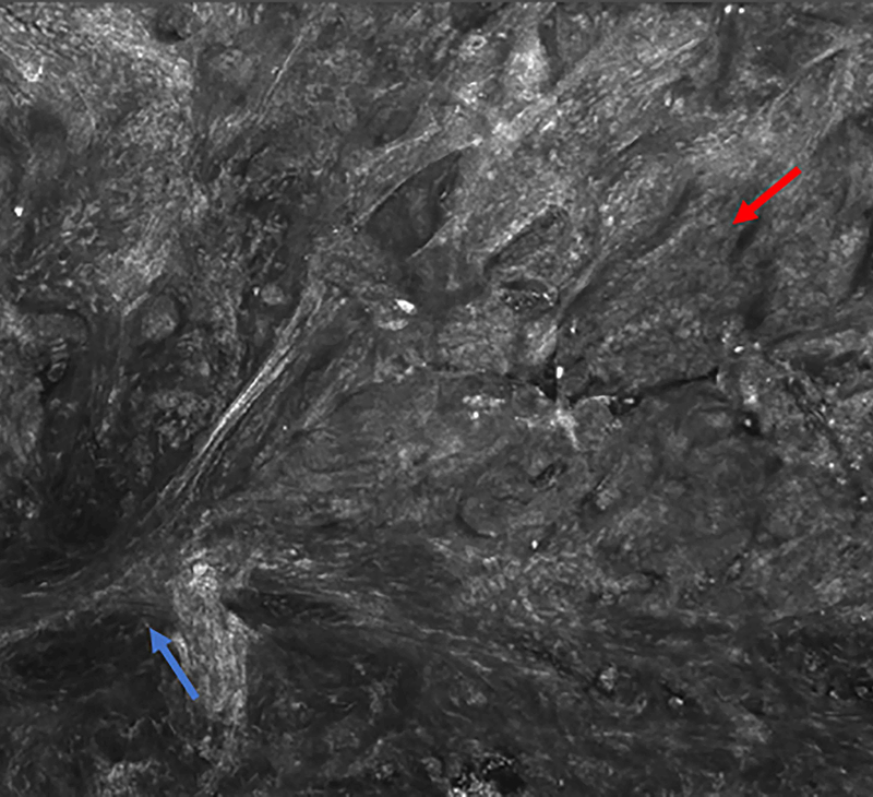
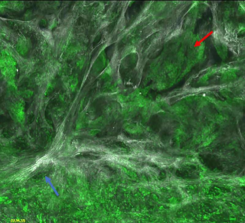
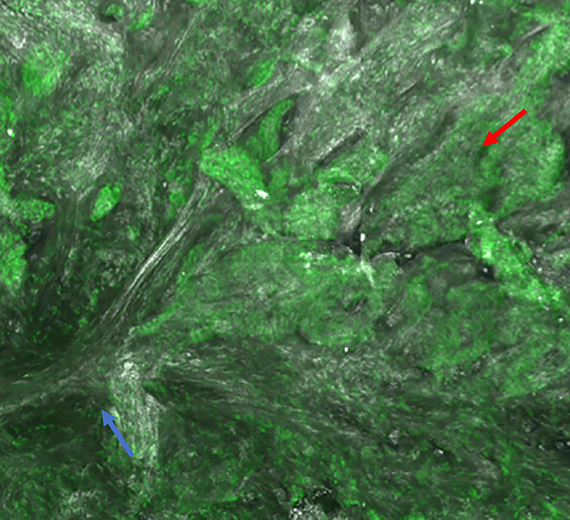
Background: Ex vivo confocal microscopy (CM) works under two modes, fluorescence and reflectance, allowing the visualization of different structures. Fluorescence CM (FCM) requires a contrast agent and has been used for the analysis of basal cell carcinomas (BCCs) during Mohs surgery. Conversely, reflectance CM (RCM) is mostly used for in vivo diagnosis of equivocal skin tumours. Recently, a new, faster ex vivo confocal microscope has been developed which simultaneously uses both lasers (fusion mode).

Objectives: To describe the BCC features identified on reflectance, fluorescence and fusion modes using this novel device. To determine the best mode to identify characteristic BCC features. To develop a new staining protocol to improve the visualization of BCC under the different modes.

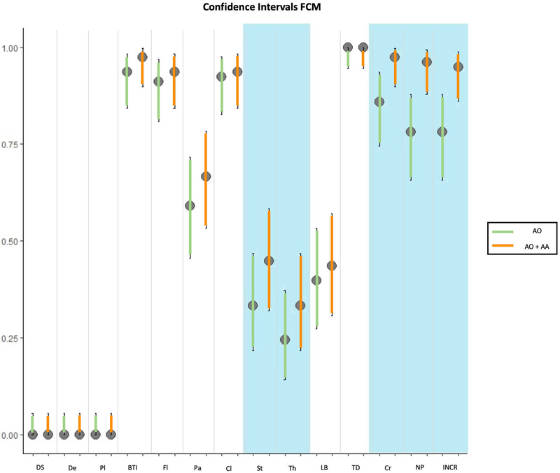
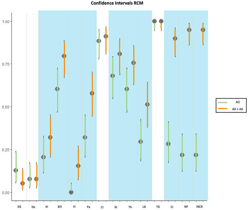
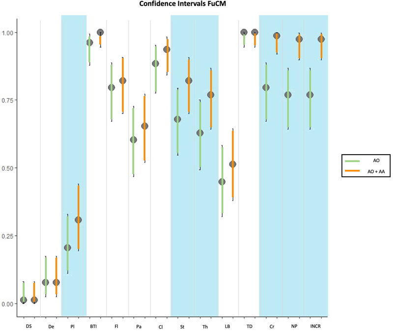
Methods: From September 2016 to June 2017, we prospectively included consecutive BCCs which were excised using Mohs surgery in our department. The lesions were evaluated using ex vivo CM after routine Mohs surgery. The specimens were first stained with acridine orange and then stained using both acetic acid and acridine orange.

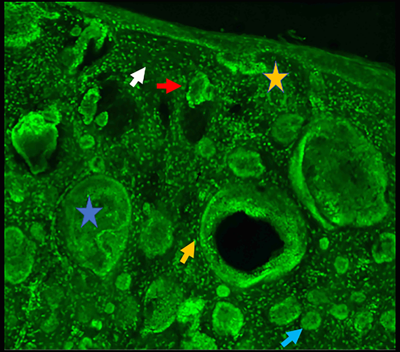
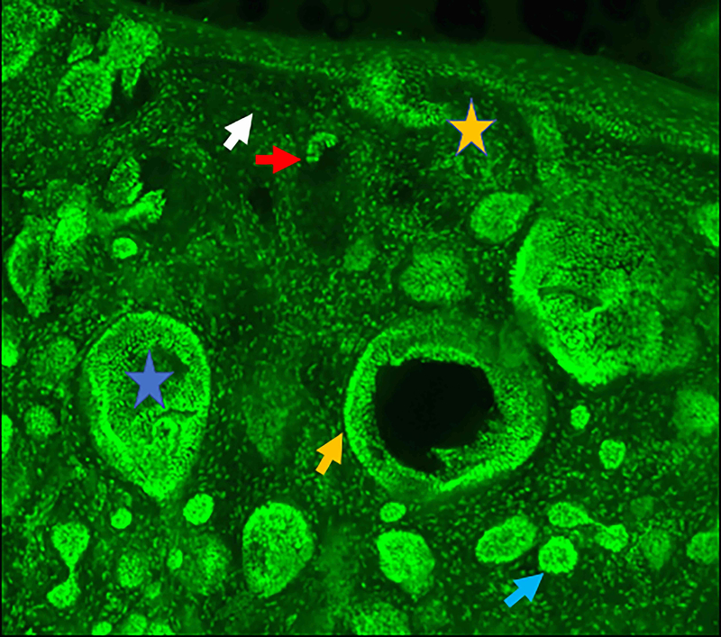
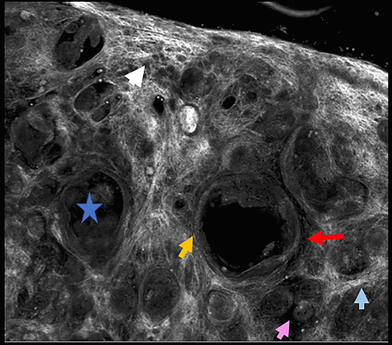
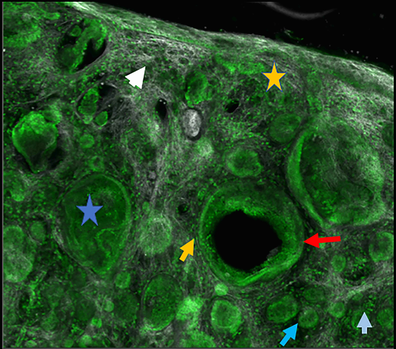
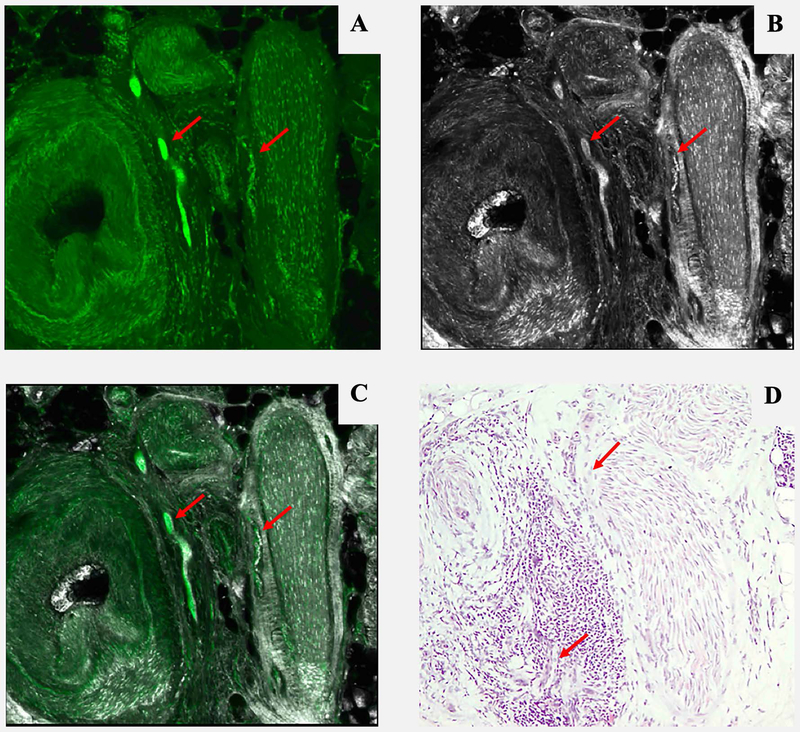
Results: We included 78 BCCs (35 infiltrative, 25 nodular, 12 micronodular, 6 superficial). Most features were better visualized with the fusion mode using the double staining. We also identified new CM ex vivo features, dendritic and plump cells, which have not been reported previously.

Conclusions: Our results suggest that nuclei characteristics are better visualized in FCM but cytoplasm and surrounding stroma are better visualized in RCM. Thus, the simultaneous evaluation of reflectance and fluorescence seems to be beneficial due to its complementary effect. What's already known about this topic? Ex vivo fluorescent confocal microscopy (FCM) is an imaging technique that allows histopathological analysis of fresh tissue. FCM is faster - at least one-third of the time - than conventional methods. FCM has a sensitivity of 88% and a specificity of 99% in detecting basal cell carcinomas (BCCs). What does this study add? Reflectance and fluorescence modes can be used simultaneously in a new ex vivo CM device. Each mode complements the other, resulting in an increase in the detection of BCC features in fusion mode. A combined staining using acetic acid and acridine orange enhances the visualization of tumour and stroma without damaging the tissue for further histopathological analysis.


Conflict of interest statement

Disclosures: The authors have no disclosures or conflicts of interest to report.

Figures

Figure 1:
Figure 1:
Confidence intervals between different laser and staining procedures. 1A: comparison of staining in fluorescent confocal microscopy (FCM); 1B: comparison of staining in reflectance confocal microscopy (RCM); 1C: comparison of staining in fusion mode of confocal microscopy (FuCM); 1D comparison between different laser modes with the best staining (AO+AA). Grey colour for significant comparisons for p-McNemar.
Figure 1:
Figure 1:
Confidence intervals between different laser and staining procedures. 1A: comparison of staining in fluorescent confocal microscopy (FCM); 1B: comparison of staining in reflectance confocal microscopy (RCM); 1C: comparison of staining in fusion mode of confocal microscopy (FuCM); 1D comparison between different laser modes with the best staining (AO+AA). Grey colour for significant comparisons for p-McNemar.
Figure 2.
Figure 2.
Micronodular basal cell carcinoma. Feature differences in distinct lasers and stain protocols. Right column Acridine orange + acetic acid. Left column: Acridine orange. A-A1: Fluorescence confocal microscopy. B-B1: Reflectance confocal microscopy. C-C1: Fusion confocal microscopy. Differences between stroma reactions can be observed (white arrow); clefting (red arrow); palisading (yellow arrow); thickened collagen bundles (sky blue arrow); well delineated and bright tumour islands (blue arrow); dark silhouettes (pink arrows); presence of fluorescence (yellow star); nuclear features: crowding, nuclear pleomorphism, increased nucleus/cytoplasm ratio (blue star).
Figure 2.
Figure 2.
Micronodular basal cell carcinoma. Feature differences in distinct lasers and stain protocols. Right column Acridine orange + acetic acid. Left column: Acridine orange. A-A1: Fluorescence confocal microscopy. B-B1: Reflectance confocal microscopy. C-C1: Fusion confocal microscopy. Differences between stroma reactions can be observed (white arrow); clefting (red arrow); palisading (yellow arrow); thickened collagen bundles (sky blue arrow); well delineated and bright tumour islands (blue arrow); dark silhouettes (pink arrows); presence of fluorescence (yellow star); nuclear features: crowding, nuclear pleomorphism, increased nucleus/cytoplasm ratio (blue star).
Figure 3.
Figure 3.
Infiltrating basal cell carcinoma. Feature differences: distinct lasers and stain protocols. Right column Acridine orange + acetic acid. Left column: Acridine orange. A-A1: Fluorescence confocal microscopy. B-B1: Reflectance confocal microscopy. C-C1: Fusion confocal microscopy. Dark silhouettes are better observed without the nuclear enhancement of the acetic acid. They are poorly visualized in fluorescence mode and better observed in reflectance mode. When the acetic acid is applied, bright and well delineated tumour islands can be observed instead of them (red arrow). Collagen bundles are poorly observed in fluorescence mode (blue arrow). Clefting is not always observed in infiltrating tumours.
Figure 3.
Figure 3.
Infiltrating basal cell carcinoma. Feature differences: distinct lasers and stain protocols. Right column Acridine orange + acetic acid. Left column: Acridine orange. A-A1: Fluorescence confocal microscopy. B-B1: Reflectance confocal microscopy. C-C1: Fusion confocal microscopy. Dark silhouettes are better observed without the nuclear enhancement of the acetic acid. They are poorly visualized in fluorescence mode and better observed in reflectance mode. When the acetic acid is applied, bright and well delineated tumour islands can be observed instead of them (red arrow). Collagen bundles are poorly observed in fluorescence mode (blue arrow). Clefting is not always observed in infiltrating tumours.
Figure 4.
Figure 4.
Three-dimensional peri vascular and peri neural infiltrating basal cell carcinoma (red arrow). A: Fluorescence mode. B: Reflectance mode. C: Fusion mode. D: Hematoxylin and Eosin. 10x. Even small amounts of tumour can be detected by this method, but not always clearly by conventional pathology.

Comment in

Similar articles

Cited by

References

    1. Forkner C A method for supravital staining of animals with neutral red and its preservation in paraffin sections. J Exp Med 1930; 52:379–84. - PMC - PubMed
    1. Alarcon I, Carrera C, Palou J, et al. Impact of in vivo reflectance confocal microscopy on the number needed to treat melanoma in doubtful lesions. Br J Dermatol 2014; 170:802–8. - PMC - PubMed
    1. Pellacani G, Pepe P, Casari A, Longo C. Reflectance confocal microscopy as a second-level examination in skin oncology improves diagnostic accuracy and saves unnecessary excisions: A longitudinal prospective study. Br J Dermatol 2014; 171. doi:10.1111/bjd.13148. - DOI - PubMed
    1. Stevenson AD, Mickan S, Mallett S, Ayya M. Systematic review of diagnostic accuracy of reflectance confocal microscopy for melanoma diagnosis in patients with clinically equivocal skin lesions. Dermatol Pract Concept 2013; 3:19–27. - PMC - PubMed
    1. Bennàssar A, Vilata A, Puig S, Malvehy J. Ex vivo fluorescence confocal microscopy for fast evaluation of tumour margins during Mohs surgery. Br J Dermatol 2014; 170:360–5. - PubMed

Publication types