Review
. 2019 Sep;17(9):1430-1439.
doi: 10.1111/jth.14544. Epub 2019 Jul 16.

New insights into cytoskeletal remodeling during platelet production

Affiliations
Review

New insights into cytoskeletal remodeling during platelet production

Dorsaf Ghalloussi et al. J Thromb Haemost. 2019 Sep.

Abstract

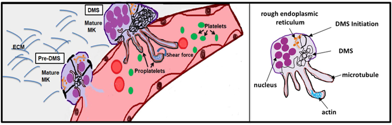
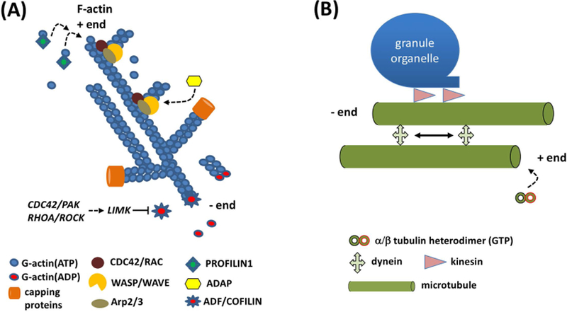
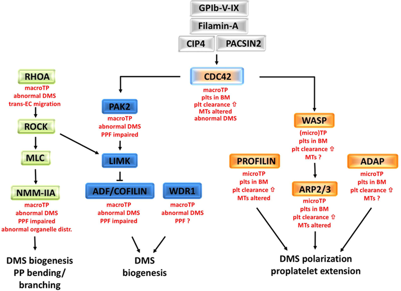
The past decade has brought unprecedented advances in our understanding of megakaryocyte (MK) biology and platelet production, processes that are strongly dependent on the cytoskeleton. Facilitated by technological innovations, such as new high-resolution imaging techniques (in vitro and in vivo) and lineage-specific gene knockout and reporter mouse strains, we are now able to visualize and characterize the molecular machinery required for MK development and proplatelet formation in live mice. Whole genome and RNA sequencing analysis of patients with rare platelet disorders, combined with targeted genetic interventions in mice, has led to the identification and characterization of numerous new genes important for MK development. Many of the genes important for proplatelet formation code for proteins that control cytoskeletal dynamics in cells, such as Rho GTPases and their downstream targets. In this review, we discuss how the final stages of MK development are controlled by the cellular cytoskeletons, and we compare changes in MK biology observed in patients and mice with mutations in cytoskeleton regulatory genes.

Keywords: bone marrow; cytoskeleton; megakaryocytes; platelets; proplatelet formation.


Conflict of interest statement

Conflicts of interest

The authors have no conflicts of interest to declare.

Figures

Figure 1
Figure 1. Schematic representation showing the final stages of MK development leading to platelet release into BM sinusoids.
Terminal MKs enter the process of proplatelet formation by establishing an expansive and interconnected membranous network, called the Demarcation Membrane System (DMS). Signals initiated at the cell membrane lead to formation of focal tubular membrane invaginations and the generation of surface-connected intracellular membrane pools, called the pre-DMS, which are precisely located along the cleavage furrow of the endomitotic MK. Maturation of the pre-DMS into the DMS is dependent on the insertion of additional membranes. Once it is fully established, the DMS provides the membranes required for the extension of proplatelet shafts, cellular protrusions that shed proplatelets from their tips. Proplatelet formation (PPF) requires dynamic remodeling and profound changes in the microtubule (MT) and actin cytoskeleton. The release of proplatelet structures, which mature into platelets, into the blood stream is supported by shear force and turbulence. ECM: extracellular matrix.
Figure 2
Figure 2. DMS biogenesis and proplatelet elongation depend on the actin and microtubule cytoskeletons.
(A) The actin cytoskeleton is critical for DMS biogenesis. Filamentous (F-) actin is organized in a branched network. Plus (+) ends grow rapidly by addition of G-actin ATP monomers, facilitated by PROFILIN1. The ARP2/3 complex, controlled by the CDC42-WASP and/or the RAC/WAVE pathways, initiates branching of existing F-actin filaments. The adapter protein, ADAP, assists in this process. Binding of capping proteins to the +ends limits the growth of F-actin filaments. Actin depolymerizing factor (ADF)/COFILIN is a family of actin-binding proteins which disassembles actin filaments. Its activity is inhibited by LIM kinase (LIMK), which is controlled by the CDC42/PAK and RHOA/ROCK pathways. (B) Proplatelet elongation is microtubule (MT)-driven. MTs are α/β tubulin heterodimer polymers, which form rigid tubes of ~25nm in diameter. MTs are polar structures that grow from their plus (+) end. Proplatelet shafts are elongated by two separate MT activities: the continuous growth of MT + ends and sliding of adjacent MTs relative to one another. Cytoplasmic dynein is a motor protein responsible for sliding of MTs relative to one another, the major mechanism underlying proplatelet growth. Kinesin is another motor protein required for cargo (organelles, granules) movement along the MTs.
Figure 3
Figure 3. Role of Rho GTPases and their downstream targets in DMS biogenesis and proplatelet formation.
Small GTPases are important regulators of the MK cytoskeletons, DMS biogenesis, and proplatelet formation. Activation of CDC42 is dependent on the GPIb-V-IX complex, filamin A, and the F-BAR proteins, CIP4 and PACSIN2 (grey boxes). CDC42 affects the activity state of ADF/COFILIN via PAK2/LIMK signaling for proper DMS formation (blue boxes). WD40 repeat protein 1 (WDR1) forms a complex with COFILIN. Downstream of RHOA, ROCK controls F-actin levels via ADF/COFILIN and bending via MLC and non-muscle myosin (NMM-IIA) (green boxes). Macrothrombocytopenia (macroTP), an abnormal DMS, and impaired PPF are common to mutant mice with defects in RHOA/ROCK or CDC42/PAK2 signaling pathways. However, mice deficient in CDC42 show additional defects such as ectopic platelet release into the bone marrow niche and increased clearance of platelets from the circulation. Similar defects are observed in mice deficient in WASP, ARP2/3, PROFILIN1, and ADAP (orange boxes), suggesting that these proteins are part of the same signaling pathway. Micro-thrombocytopenia (microTP), an altered tubulin cytoskeleton in platelets, abnormal DMS polarization, and defective proplatelet extension are additional defects characteristic for these mice. The small GTPase RAC1 can partially compensate for deficiency in CDC42 (not shown). Listed in red font are the main MK and platelet phenotypes in mice with defects in the respective signaling molecules.

Similar articles

Cited by

References

    1. Tomaiuolo M, Brass LF, Stalker TJ. Regulation of Platelet Activation and Coagulation and Its Role in Vascular Injury and Arterial Thrombosis. Interventional Cardiology Clinics 2017. - PMC - PubMed
    1. Boulaftali Y, Hess PR, Getz TM, Cholka A, Stolla M, Mackman N, Owens AP, Ware J, Kahn ML, Bergmeier W. Platelet ITAM signaling is critical for vascular integrity in infammation. J Clin Invest 2013; 123: 908–16. - PMC - PubMed
    1. Grozovsky R, Giannini S, Falet H, Hoffmeister KM. Regulating billions of blood platelets: Glycans and beyond. Blood 2015. - PMC - PubMed
    1. Kaufman RM, Airo R, Pollack S, Crosby WH. Circulating megakaryocytes and platelet release in the lung. Blood 1965; 26(6): 720–31. - PubMed
    1. Lefrançais E, Ortiz-Muñoz G, Caudrillier A, Mallavia B, Liu F, Sayah DM, Thornton EE, Headley MB, David T, Coughlin SR, Krummel MF, Leavitt AD, Passegué E, Looney MR. The lung is a site of platelet biogenesis and a reservoir for haematopoietic progenitors. Nature 2017; 544(7648): 105–9. - PMC - PubMed

Publication types