Review
. 2019 Jun 20:88:163-190.
doi: 10.1146/annurev-biochem-013118-110644.

Redox Chemistry in the Genome: Emergence of the [4Fe4S] Cofactor in Repair and Replication

Affiliations
Review

Redox Chemistry in the Genome: Emergence of the [4Fe4S] Cofactor in Repair and Replication

Jacqueline K Barton et al. Annu Rev Biochem. .

Abstract

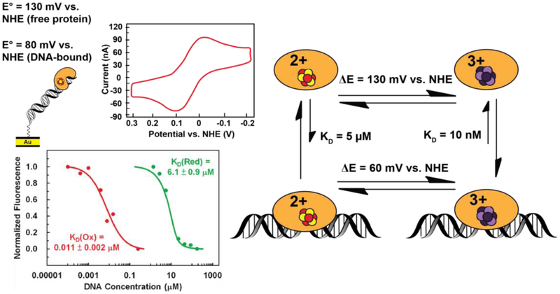
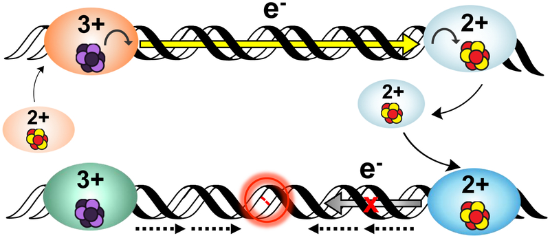
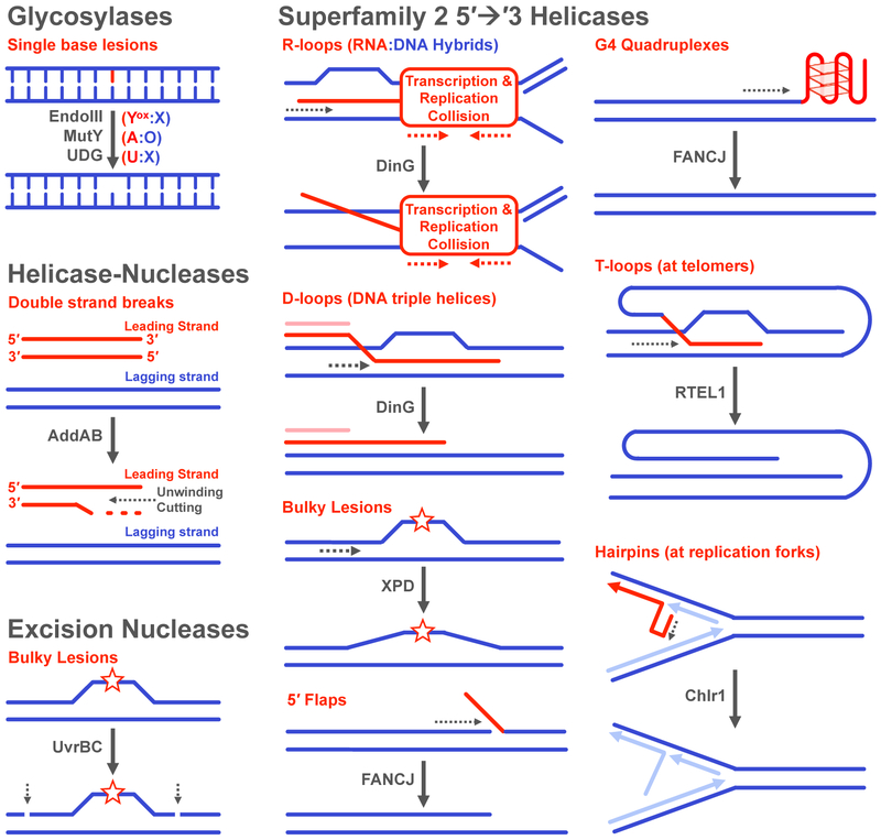
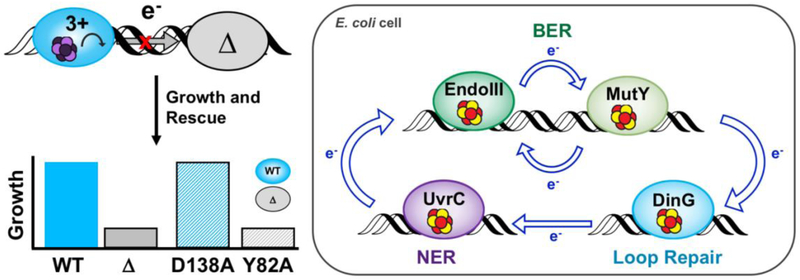
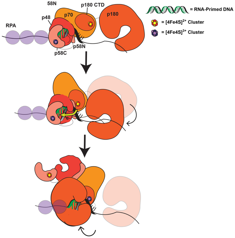
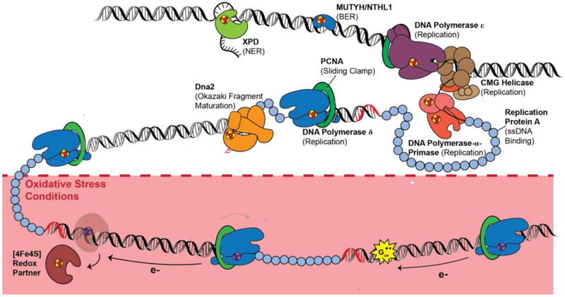
Many DNA-processing enzymes have been shown to contain a [4Fe4S] cluster, a common redox cofactor in biology. Using DNA electrochemistry, we find that binding of the DNA polyanion promotes a negative shift in [4Fe4S] cluster potential, which corresponds thermodynamically to a ∼500-fold increase in DNA-binding affinity for the oxidized [4Fe4S]3+ cluster versus the reduced [4Fe4S]2+ cluster. This redox switch can be activated from a distance using DNA charge transport (DNA CT) chemistry. DNA-processing proteins containing the [4Fe4S] cluster are enumerated, with possible roles for the redox switch highlighted. A model is described where repair proteins may signal one another using DNA-mediated charge transport as a first step in their search for lesions. The redox switch in eukaryotic DNA primases appears to regulate polymerase handoff, and in DNA polymerase δ, the redox switch provides a means to modulate replication in response to oxidative stress. We thus describe redox signaling interactions of DNA-processing [4Fe4S] enzymes, as well as the most interesting potential players to consider in delineating new DNA-mediated redox signaling networks.

Keywords: DNA charge transport; DNA polymerase; DNA primase; base excision repair; iron–sulfur clusters; oxidative stress; redox signaling.


Figures

Figure 1.
Figure 1.. [4Fe4S] Clusters.
(Above, left) Schematic of the cubane [4Fe4S] cluster, with Fe in red and S in yellow. (Above, right) Cofactors containing iron can react with cellular oxidants leading to Fenton chemistry and DNA damage by the hydroxyl radical. (Below) The potential of the [4Fe4S] cluster cofactor is tunable over a wide range of physiological redox potential values. Ferredoxins access the [4Fe4S]2+/1+ couple, upon reduction from the resting [4Fe4S]2+ state. (yellow). High potential iron proteins (HiPIPs) access the [4Fe4S]3+/2+ couple upon oxidation to the [4Fe4S]3+ state from the resting [4Fe4S]2+ state (red). DNA-processing enzymes with [4Fe4S] cofactors have DNA-bound redox potentials which fall within the HiPIP [4Fe4S]3+/2+ potential range, at approximately ~65-150mV vs. NHE (blue). The solvent accessibility and hydrogen bonding/electrostatic environment of the cluster all contribute to tuning the redox potential of the cofactor (4,5).
Figure 2.
Figure 2.. [4Fe4S] cluster biogenesis and loading into prokaryotic (above) and eukaryotic (below) target proteins.
Scaffold proteins, together with cysteine desulfurases and ferrous iron sources, assemble the cluster and bind chaperone machinery to then transport the cofactor first to machinery responsible for cluster delivery, then finally to target proteins. This process is not identical in bacteria and eukaryotes. However each process requires the concerted action of several specialized proteins and requires significant metabolic expense for the cell (13-19).
Figure 3.
Figure 3.. The structure of DNA facilitates long-range, rapid electron transfer.
(Top, left) Side view of DNA. The aromatic bases at the center of the DNA helix are oriented so that the π orbitals of adjacent bases overlap with one another in the duplex. This structural property suggests that charge could pass through the π-stacked base pairs of DNA. (Top, right) View down the helical axis of aromatic base pairs (blue) stacked in 3.4 Å layers at the center of DNA. (Bottom) Time scale and length scale of various electron transfer pathways through biomolecules (10, 11,41,43). PDB ID 3BSE.
Figure 4.
Figure 4.. Measuring the redox potential of DNA-bound [4Fe4S] enzymes.
(Top) DNA electrochemistry on Au electrodes, where DNA duplexes containing a tethered alkanethiol are attached to a gold electrode passivated with β-mercaptohexanol, can be used to assess redox signals from DNA-bound, [4Fe4S] proteins (yellow, left). Signals are attenuated when base lesions (red, center) in the duplex are present, or when the redox pathway within the [4Fe4S] protein, as for Y82A (gray, right) is deficient in CT. Cyclic voltammetry (below, left) scanning can be used to measure the DNA-mediated redox signal from CT-proficient proteins (WT EndoIII, blue) and CT-deficient proteins (EndoIII Y82A, red). A multiplexed chip platform (below, right) has now been adapted to measure [4Fe4S] protein signals on 16 separate DNA-modified electrodes, with replicates on a single surface (32, 48-50).
Figure 5.
Figure 5.. DNA binding shifts the potential of [4Fe4S] cluster enzymes.
Endonuclease III has a redox potential of approximately 80mV vs. NHE for the [4Fe4S]3+/2+ couple measured on a DNA-modified Au electrode. (Above, left) This potential is a negative shift from the ~130mV vs. NHE potential for this couple when the protein is not bound to DNA. This shift corresponds thermodynamically to a stabilization of the oxidized [4Fe4S]3+ state upon binding the DNA polyanion (right). Microscale thermophoresis on electrochemically oxidized and native reduced Endonuclease III (below, left) indicates that a ~550-fold increase in DNA binding affinity is associated with the conversion from [4Fe4S]2+ to the [4Fe4S]3+ state, (53) consistent with this negative shift in potential.
Figure 6.
Figure 6.. A model for DNA-mediated redox signaling between repair proteins.
Enzymes with the cluster in the native [4Fe4S]2+ state first bind DNA, causing the cluster to become activated toward oxidation. Oxidative stress initiates the damage search when highly reactive species such as the guanine radical cation are formed; these can oxidize DNA-bound proteins in their vicinity. Oxidation of the cluster to the [4Fe4S]3+ state leads to a > 500-fold increase in DNA binding affinity, so oxidized proteins remain bound and diffuse along the DNA. Another [4Fe4S]2+ protein bound at a distant site can reduce the oxidized protein, effectively scanning the intervening DNA for lesions through DNA-mediated CT. At this point, on damage-free DNA (above) the reduced protein binds less tightly to DNA and can diffuse away, while the newly oxidized protein continues the damage search. This process of redox exchange continues until a segment of DNA containing a lesion is approached. Since even subtle lesions can disrupt base stacking (below), CT is attenuated and any nearby oxidized proteins remain bound. Thus, DNA CT allows repair proteins to scan large sections of the genome and redistribute to areas containing damage (24, 37).
Figure 7.
Figure 7.. Reactions and Substrates for DNA repair enzymes containing [4Fe4S] clusters.
(Top, left) Glycosylases remove a number of single-base lesions caused by endogenous and exogenous agents. (Middle, left) The helicase-nuclease AddAB processes double strand breaks. (Bottom, left) The UvrBC complex cleaves the phosphodiester backbone around the damaged strand of DNA. (Right) Superfamily 2 5′→3′ helicases participate in a number of pathways and are involved in unwinding very diverse substrates. The substrate specificity is overlapping for many of the helicases (ex. FANCJ and RTEL1), though genome location (ex. telomeres) and cell cycle phase (ex. replication in the S phase) appear to be factors in activity (73, 77, 81, 82, 87, 91,93).
Figure 8.
Figure 8.. Visualization of Protein Localization on Damaged DNA by Atomic Force Microscopy (AFM).
In the AFM redistribution assay, [4Fe4S] protein or protein mixtures are incubated with DNA substrates which contain either long, well matched DNA strands (3.8 kb) or strands containing a single C:A mismatch in the 3.8 kb duplex along with short, undamaged DNA strands (1.6 and 2.2 kb). Images are collected and proteins bound to long strands of DNA are counted, normalized to the proteins bound to the short strands, and expressed as a binding density ratio (right). As shown at right (top), a greater density of proteins is found on the strands containing a mismatch (C:A) compared to the well-matched (T:A) strand, even though the repair proteins do not bind the C:A mismatch as a substrate. If the protein is defective in carrying out DNA CT, however, the binding density is the same on the mismatched and matched strands (bottom right) (55, 59, 79, 80).
Figure 9.
Figure 9.. Genetic Assays for Detection of DNACT Signaling Among E. coli [4Fe4S] Repair Proteins.
(Top left) E. coli parent (blue) and EndoIII knockout (KO, gray) strains that report on the repair protein activity have been used to monitor DNA-mediated communication between putative signaling partners in vivo. Complementation plasmids expressing CT-proficient (D138A) or CT-deficient (Y82A) versions of EndoIII are introduced to EndoIII KO strains to evaluate if the parent phenotype can be rescued (bottom left). Rescue can only be achieved with a CT-proficient enzyme (blue bar, gray outline), strongly indicating that DNA-mediated redox signaling is necessary for efficient repair. (Right) The redox signaling network in E. coli includes BER, loop repair, and NER (57, 59, 79, 80).
Figure 10.
Figure 10.. Model for primase-polymerase α handoff through redox switching.
(52) Oxidized [4Fe4S]3+ primase is bound to the RNA/DNA primer during primer synthesis. Polymerase α is DNA-dissociated and reduced but flexibly tethered to primase (Top). When the RNA primer reaches appropriate length, polymerase α orients in a manner coupling the [4Fe4S] cluster into the RNA/DNA substrate and can be oxidized by DNA CT through this segment, sending an electron through the primed template to reduce DNA primase (Middle). Reduced primase dissociates from the RNA-primed DNA, and oxidized polymerase α can then synthesize DNA downstream of the primase product (Bottom).
Figure 11.
Figure 11.. [4Fe4S] enzymes in eukaryotic repair and replication.
B-family polymerases, DNA primase, Dna2 helicase-nuclease, BER/NER enzymes such as MUTYH, NTHL1, and XPD, all coordinate a [4Fe4S] cluster cofactor. Several of these proteins have been demonstrated to participate in DNA-mediated redox signaling; characterization of their redox roles is ongoing. (Below) Under oxidative stress conditions, polymerase δ may be converted to the [4Fe4S]3+ state as a means to stall synthesis under poor cellular conditions. Polymerase δ can be reversibly oxidized and reduced through DNA CT, which may regulate polymerase activity on the lagging strand.

Similar articles

Cited by

References

    1. Beinert H, Holm RH, Munck E. 1997. Iron-sulfur clusters: Nature’s modular, multipurpose structures. Science 277:653–59 - PubMed
    1. Rees DC, Howard JB. 2003. The interface between the biological and inorganic worlds: Iron-sulfur metalloclusters. Science 300:929–31 - PubMed
    1. Meyer J 2008. Iron-sulfur protein folds, iron-sulfur chemistry, and evolution. J. Biol. Inorg. Chem 13:157–70 - PubMed
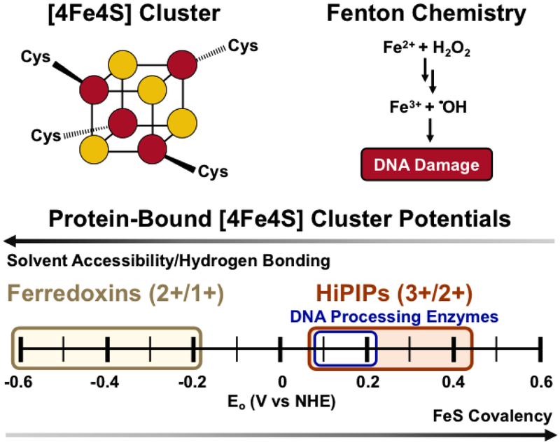
    1. Dey A, Jenney FE, Adams MWW, Babini E, Takahashi Y, et al. 2007. Solvent tuning of electrochemical potentials in the active sites of HiPIP versus ferredoxin. Science 318:1464–68 - PubMed
    1. Ha Y, Arnold AR, Nuñez NN, Bartels PL, Zhou A, et al. 2017. Sulfur K-Edge XAS Studies of the Effect of DNA Binding on the [Fe4S4] Site in EndoIII and MutY. J. Am. Chem. Soc 139:11434–42 - PMC - PubMed

Publication types

MeSH terms

LinkOut - more resources