Review
. 2019 Jun 20:88:137-162.
doi: 10.1146/annurev-biochem-013118-111315.

Eukaryotic Base Excision Repair: New Approaches Shine Light on Mechanism

Affiliations
Review

Eukaryotic Base Excision Repair: New Approaches Shine Light on Mechanism

William A Beard et al. Annu Rev Biochem. .

Abstract

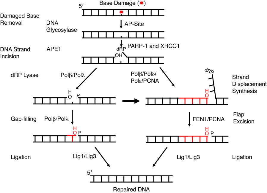
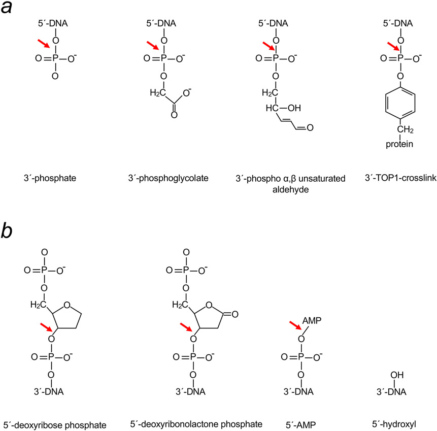
Genomic DNA is susceptible to endogenous and environmental stresses that modify DNA structure and its coding potential. Correspondingly, cells have evolved intricate DNA repair systems to deter changes to their genetic material. Base excision DNA repair involves a number of enzymes and protein cofactors that hasten repair of damaged DNA bases. Recent advances have identified macromolecular complexes that assemble at the DNA lesion and mediate repair. The repair of base lesions generally requires five enzymatic activities: glycosylase, endonuclease, lyase, polymerase, and ligase. The protein cofactors and mechanisms for coordinating the sequential enzymatic steps of repair are being revealed through a range of experimental approaches. We discuss the enzymes and protein cofactors involved in eukaryotic base excision repair, emphasizing the challenge of integrating findings from multiple methodologies. The results provide an opportunity to assimilate biochemical findings with cell-based assays to uncover new insights into this deceptively complex repair pathway.

Keywords: DNA polymerase; genome stability; mechanism; mutagenesis; repair; structure.


Figures

Figure 1
Figure 1
A simplified scheme illustrating steps and factors in canonical sub-pathways of mammalian BER. BER is initiated by DNA glycosylase removal of a damaged base or by spontaneous base loss (not shown). Following APE1 incision 5′ to the AP site sugar, repair proceeds by short-patch BER (left path) or long-patch BER (right path). A red star depicts the damaged base and nascent nucleotide(s) is shown in red.
Figure 2
Figure 2
Common BER intermediates found in the incised DNA strand that need to be processed to accomplish DNA repair. Incision of an AP-site can result in (a) a 3´-blocking group that must be removed by an enzyme to generate a 3´-hydroxyl (red arrow) or (b) a 5´-blocking group that must be removed by an enzyme to generate a 5´-phosphate (red arrow). In the case where there is a 5´-hydroxyl, PNPK can add a phosphate group to allow ligation.
Figure 3
Figure 3
Features of a high-resolution crystal structure of the human APE1 (36). (a) The structure of a substrate complex reveals that the AP-site sugar (tetrahydrofuran, THF; gray carbons) is flipped outside of the DNA helix into the APE1 (purple ribbons) active site (PDB ID: 5DFI). The DNA strand that will be incised is colored yellow and the complementary strand is green. (b) Detailed view of the active site of a product complex illustrating a magnesium (green sphere; coordination illustrated with green dashed lines) associated interaction network (black dashed lines) that includes waters (small red spheres), protein side chains (purple carbons), and product DNA (green semi-transparent bonds except the nucleotide 5´ to the gap, solid green bonds) results in tight product binding (PDB ID: 5DFI).
Figure 4
Figure 4
DNA polymerase ternary substrate complex structure (52). (a) Ribbon representation of human pol β bound to single-nucleotide gapped DNA. The template strand is gray and the primer and downstream DNA strands are yellow and the 5´-terminus of the primer strand is indicated. The amino-terminal lyase domain is red and the polymerase domain is black. (b) An alternate view of the complex highlighting HhH motifs (solid ribbon representation; residues 55–79 and 91–118 of the lyase and polymerase domains, respectively) that interact with the DNA backbone of the incised strand through Na+ ions (purple spheres). The polymerase active site Mg2+ are indicated with green spheres. The 3´-terminus of the template strand is indicated.
Figure 5
Figure 5
Electrostatic surface potential of X-family DNA polymerases. The surface potential is mapped onto the protein surface (red to blue represents negative to positive potential, ±10 kcal/mol*e). The view is similar to that in Figure 3a. A black dashed oval identifies the position of the downstream 5´-phosphate. (a) pol β (PDB ID: 2FMS) (52), (b) pol λ (PDB ID: 1XSN) (53), (c) pol μ (PDB ID: 2IHM) (51), and (d) TdT (PDB ID: 5D49) (54). The DNA template strand is colored gray and primer and downstream strands are colored yellow.
Figure 6
Figure 6
Sucrose density gradient centrifugation analyses of a large BER complex. (a) A BER complex isolated by affinity-capture chromatography with pol β as the bait was analyzed, and gradient fractions were probed by immunoblotting with anti-pol β antibody. The migration positions of the large BER complex and monomeric pol β are shown. (b) The results of re-centrifugation of the large BER complex using a more dense gradient than in (a). (c) Sucrose gradient analysis of the crude cell extract prior to affinity-capture chromatography; gradient fractions were probed by immunoblotting with the three antibodies shown. APE1 was not found in the gradient region containing the large BER complex and only a minor portion of pol β and PARP-1 were found in the large complex. Reproduced from Figure 2 (61).
Figure 7
Figure 7
Time course of recruitment of transiently expressed carboxyl-terminal tagged repair proteins. Wild-type XRCC1 cells were subjected to micro-irradiation damage and recruitment followed for 200 s. (a) Typical accumulation of fluorescently tagged proteins and enhanced green fluorescent protein (eGFP) at damaged sites after 100 s repair. Recruitment curves for (b) PARP-1-YFP (YFP, yellow fluorescent protein) (c) XRCC1-GFP (d) PNKP-GFP and (e) Tdp1-GFP. Error bars represent standard error of the mean. Fluorescence data were normalized using the intensity at the beginning of recruitment and maximal intensity values, and recruitment kinetics were fitted to single exponentials except for PARP-1-YFP that was fitted to two exponentials. The recruitment half-times are given in the appropriate panel. Adapted from Figure 2 (96).
Figure 8
Figure 8
Immunofluorescent imaging of XRCC1, PAR and pol β. Cells were laser micro-irradiated in stripes to initiate XRCC1 and pol β recruitment and synthesis of PAR. After 1 min, cells were fixed and stained and a comparison of representative XC5 (XRCC1 wild-type), XCKD16 (XRCC1 mutated to prevent interaction with PNKP) and Xrcc1−/− cells is shown. Adapted from Figure 4 (96).

Similar articles

Cited by

References

    1. Sancar A 2003. Structure and function of DNA photolyase and cryptochrome blue-light photoreceptors. Chem. Rev 103: 2203–38 - PubMed
    1. Sancar A 2004. In Adv. Protein Chem, pp. 73–100: Academic Press - PubMed
    1. Lindahl T 1982. DNA repair enzymes. Ann. Rev. Biochem 51: 61–87 - PubMed
    1. Lindahl T, Demple B, Robins P. 1982. Suicide inactivation of the E. coli O6-methylguanine-DNA methyltransferase. EMBO J. 1: 1359–63 - PMC - PubMed
    1. Mitra S, Pal BC, Foote RS. 1982. O6-methylguanine-DNA methyltransferase in wild-type and ada mutants of Escherichia coli. J. Bacteriol 152: 534–37 - PMC - PubMed

Publication types

MeSH terms