

Concentration, distribution, and influence of aging on the 18 kDa translocator protein in human brain: Implications for brain imaging studies
- PMID: 31220997
- PMCID: PMC7181090
- DOI: 10.1177/0271678X19858003
Concentration, distribution, and influence of aging on the 18 kDa translocator protein in human brain: Implications for brain imaging studies
Abstract
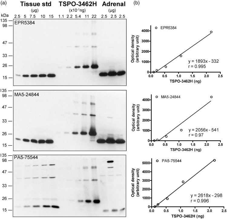
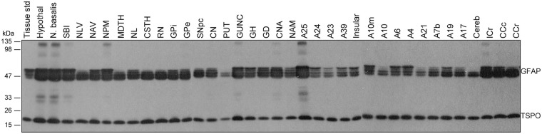
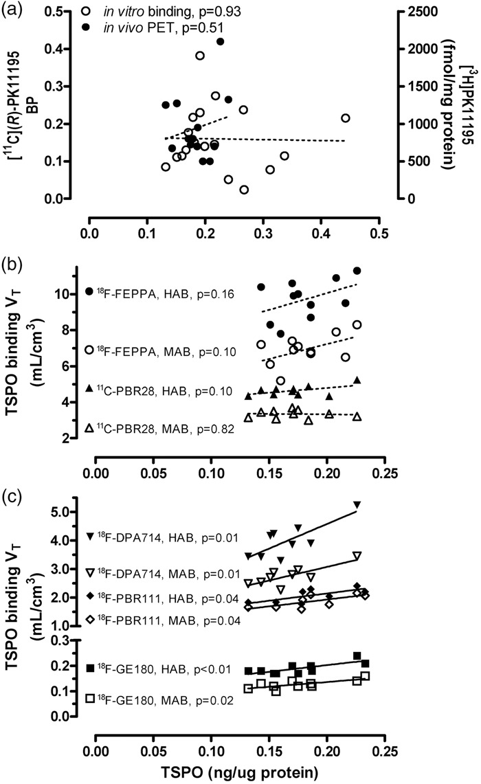
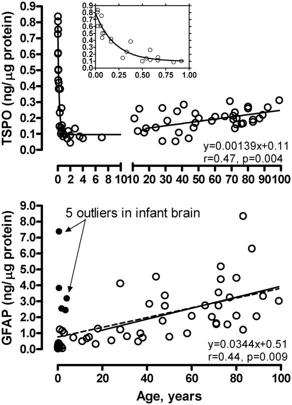
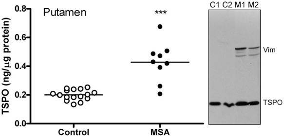
Positron emission tomography (PET) imaging of the translocator protein (TSPO) is widely used as a biomarker of microglial activation. However, TSPO protein concentration in human brain has not been optimally quantified nor has its regional distribution been compared to TSPO binding. We determined TSPO protein concentration, change with age, and regional distribution by quantitative immunoblotting in autopsied human brain. Brain TSPO protein concentration (>0.1 ng/µg protein) was higher than those reported by in vitro binding assays by at least 2 to 70 fold. TSPO protein distributed widely in both gray and white matter regions, with distribution in major gray matter areas ranked generally similar to that of PET binding in second-generation radiotracer studies. TSPO protein concentration in frontal cortex was high at birth, declined precipitously during the first three months, and increased modestly during adulthood/senescence (10%/decade; vs. 30% for comparison astrocytic marker GFAP). As expected, TSPO protein levels were significantly increased (+114%) in degenerating putamen in multiple system atrophy, providing further circumstantial support for TSPO as a gliosis marker. Overall, findings show some similarities between TSPO protein and PET binding characteristics in the human brain but also suggest that part of the TSPO protein pool might be less available for radioligand binding.
Keywords: Translocator protein TSPO; aging; multiple system atrophy; positron emission tomography; postmortem human brain.
Figures
Similar articles
-
Imaging brain microglial activation using positron emission tomography and translocator protein-specific radioligands.Int Rev Neurobiol. 2011;101:19-39. doi: 10.1016/B978-0-12-387718-5.00002-X. Int Rev Neurobiol. 2011. PMID: 22050847 Review.
-
Upregulation of cannabinoid receptor type 2, but not TSPO, in senescence-accelerated neuroinflammation in mice: a positron emission tomography study.J Neuroinflammation. 2019 Nov 10;16(1):208. doi: 10.1186/s12974-019-1604-3. J Neuroinflammation. 2019. PMID: 31707986 Free PMC article.
-
Age and disease related changes in the translocator protein (TSPO) system in the human brain: positron emission tomography measurements with [11C]vinpocetine.Neuroimage. 2011 Jun 1;56(3):1111-21. doi: 10.1016/j.neuroimage.2011.02.020. Epub 2011 Feb 12. Neuroimage. 2011. PMID: 21320609
-
Assessment of simplified methods for quantification of [18F]-DPA-714 using 3D whole-brain TSPO immunohistochemistry in a non-human primate.J Cereb Blood Flow Metab. 2020 May;40(5):1103-1116. doi: 10.1177/0271678X19859034. Epub 2019 Jun 25. J Cereb Blood Flow Metab. 2020. PMID: 31238764 Free PMC article.
-
Radiolabelled molecules for imaging the translocator protein (18 kDa) using positron emission tomography.Curr Med Chem. 2009;16(22):2899-923. doi: 10.2174/092986709788803150. Curr Med Chem. 2009. PMID: 19689272 Review.
Cited by
-
Microglia imaging in methamphetamine use disorder: a positron emission tomography study with the 18 kDa translocator protein radioligand [F-18]FEPPA.Addict Biol. 2021 Jan;26(1):e12876. doi: 10.1111/adb.12876. Epub 2020 Feb 4. Addict Biol. 2021. PMID: 32017280 Free PMC article.
-
18F-Radiolabeled Translocator Protein (TSPO) PET Tracers: Recent Development of TSPO Radioligands and Their Application to PET Study.Pharmaceutics. 2022 Nov 21;14(11):2545. doi: 10.3390/pharmaceutics14112545. Pharmaceutics. 2022. PMID: 36432736 Free PMC article. Review.
-
TSPO PET detects acute neuroinflammation but not diffuse chronically activated MHCII microglia in the rat.EJNMMI Res. 2020 Sep 29;10(1):113. doi: 10.1186/s13550-020-00699-x. EJNMMI Res. 2020. PMID: 32990808 Free PMC article.
-
Towards PET imaging of the dynamic phenotypes of microglia.Clin Exp Immunol. 2021 Dec;206(3):282-300. doi: 10.1111/cei.13649. Epub 2021 Aug 16. Clin Exp Immunol. 2021. PMID: 34331705 Free PMC article. Review.
-
Hemispheric asymmetry of [11C](R)PK11195 binding to translocator protein 18 kDa (TSPO) in normal brain.J Cereb Blood Flow Metab. 2025 Jun 19:271678X251348790. doi: 10.1177/0271678X251348790. Online ahead of print. J Cereb Blood Flow Metab. 2025. PMID: 40536168 Free PMC article.
References
-
- Papadopoulos V, Baraldi M, Guilarte TR, et al.Translocator protein (18kDa): new nomenclature for the peripheral-type benzodiazepine receptor based on its structure and molecular function. Trends Pharmacol Sci 2006; 27: 402–409. - PubMed
-
- Braestrup C, Albrechtsen R, Squires RF. High densities of benzodiazepine receptors in human cortical areas. Nature 1977; 269: 702–704. - PubMed
-
- Schoemaker H, Bliss M, Yamamura HI. Specific high-affinity saturable binding of [3H] R05-4864 to benzodiazepine binding sites in the rat cerebral cortex. Eur J Pharmacol 1981; 71: 173–175. - PubMed
-
- Benavides J, Quarteronet D, Imbault F, et al.Labelling of “peripheral-type” benzodiazepine binding sites in the rat brain by using [3H]PK 11195, an isoquinoline carboxamide derivative: kinetic studies and autoradiographic localization. J Neurochem 1983; 41: 1744–1750. - PubMed
-
- Schoemaker H, Morelli M, Deshmukh P, et al.[3H]Ro5-4864 benzodiazepine binding in the kainate lesioned striatum and Huntington’s diseased basal ganglia. Brain Res 1982; 248: 396–401. - PubMed
Publication types
MeSH terms
Substances
LinkOut - more resources
Full Text Sources
Medical
Research Materials
Miscellaneous
