. 2019 Jul 9;116(28):13943-13951.
doi: 10.1073/pnas.1903297116. Epub 2019 Jun 20.

A structure-based mechanism of cisplatin resistance mediated by glutathione transferase P1-1

Affiliations

A structure-based mechanism of cisplatin resistance mediated by glutathione transferase P1-1

Anastasia De Luca et al. Proc Natl Acad Sci U S A. .

Abstract

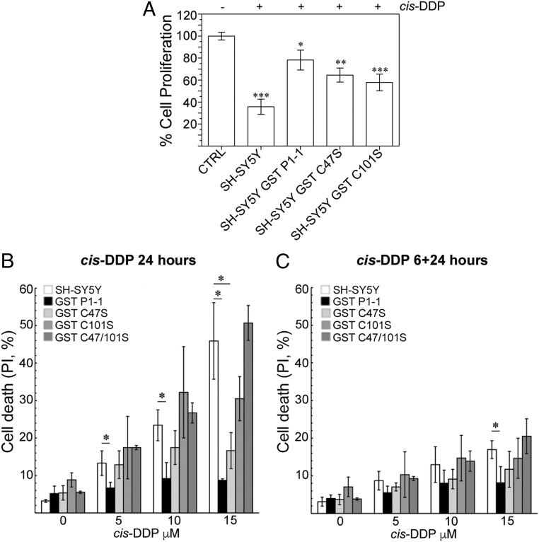
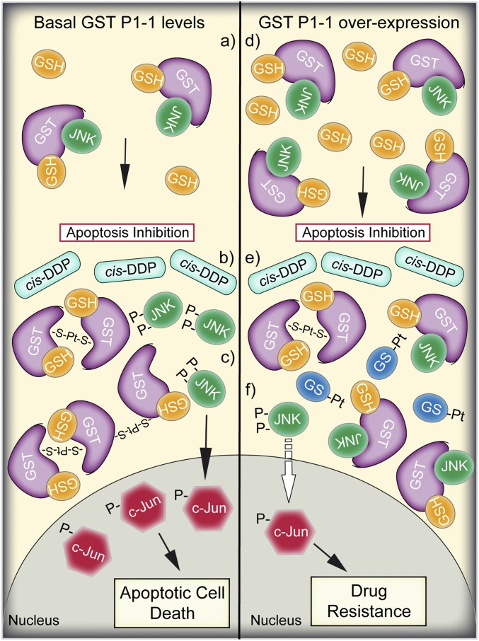
Cisplatin [cis-diamminedichloroplatinum(II) (cis-DDP)] is one of the most successful anticancer agents effective against a wide range of solid tumors. However, its use is restricted by side effects and/or by intrinsic or acquired drug resistance. Here, we probed the role of glutathione transferase (GST) P1-1, an antiapoptotic protein often overexpressed in drug-resistant tumors, as a cis-DDP-binding protein. Our results show that cis-DDP is not a substrate for the glutathione (GSH) transferase activity of GST P1-1. Instead, GST P1-1 sequesters and inactivates cisplatin with the aid of 2 solvent-accessible cysteines, resulting in protein subunits cross-linking, while maintaining its GSH-conjugation activity. Furthermore, it is well known that GST P1-1 binding to the c-Jun N-terminal kinase (JNK) inhibits JNK phosphorylation, which is required for downstream apoptosis signaling. Thus, in turn, GST P1-1 overexpression and Pt-induced subunit cross-linking could modulate JNK apoptotic signaling, further confirming the role of GST P1-1 as an antiapoptotic protein.

Keywords: cisplatin; drug resistance; glutathione transferase; protein crystallography; protein–ligand interactions.


Conflict of interest statement

The authors declare no conflict of interest.

Figures

Fig. 1.
Fig. 1.
GST P1-1 protects tumor cells against toxicity and apoptosis following cis-DDP treatment. (A) Cell viability of SH-SY5Y cell lines expressing WT and mutated GST P1-1, after cis-DDP administration. MTS assay was performed on cells treated with 5 µM cis-DDP for 24 h. Values, normalized to untreated control cells, are expressed as means ± SD from 3 independent experiments. Statistical significance compared with untreated samples (Student’s t test): *P < 0.05, **P < 0.01, ***P < 0.001. SH-SY5Y, nontransfected cells; −, cis-DDP–negative samples; +, after exposure to cis-DDP 5 µM for 24 h. (B) FACS analysis of apoptosis induction of SH-SY5Y cell lines expressing WT and mutated GST P1-1, after cis-DDP administration for 24 h. Values are expressed as means ± SD from 6 independent experiments; *P < 0.05. (C) Cytofluorometric analysis of apoptosis induction of SH-SY5Y cell lines expressing WT and mutated GST P1-1, after cis-DDP administration for 6 h followed by a recovery for additional 24 h. Values are expressed as means ± SD from 6 independent experiments; *P < 0.05.
Fig. 2.
Fig. 2.
cis-DDP–GSH adduct formation is not affected by GST P1-1 enzymatic activity. (A) UV spectral change during the reaction of cis-DDP with GSH. cis-DDP (1 mM) was incubated with GSH (2 mM) in PBS at 37 °C for 6 h. Every 10 min, the adduct formation was followed spectrophotometrically by measuring its absorbance at 260 nm. (Inset) The UV spectral change during 6 h. (B) Purification of the cis-DDP adduct of GSH. After incubation of cis-DDP with GSH, the reaction product was eluted from an anion-exchange column with 0.2 M HCl, and the elution profile was monitored at 260 nm. Peak a represents the unbound cis-DDP while peak b contains the GS-Pt adducts collected after elution with 0.2 M HCl. (C) Reaction of cis-DDP with GSH in the presence of GST P1-1. The rate of adduct formation, either in PBS or in 2 mM NaCl (pH 7.4), was followed at 260 nm at 37 °C for 10 min and reported as the change in absorbance per minute (∆A/min), in the absence or in the presence of increasing amounts of GST P1-1. (D) Time course inactivation of GST P1-1 and its mutants in the presence of cis-DDP. , GST P1-1 alone as control; △, GST P1-1 + 10 mM GSH + 1 mM cis-DDP in PBS; ●, GST P1-1 + 1 mM cis-DDP in PBS; ♦, Cys101Ser + 1 mM cis-DDP in 2 mM NaCl; ▲, Cys47Ser + 1 mM cis-DDP in 2 mM NaCl; ■, GST P1-1 + 1 mM cis-DDP in 2 mM NaCl. Data represent means ± SD of 3 independent experiments.
Fig. 3.
Fig. 3.
cis-DDP acts as an effective alkylating agent of GST P1-1, in the absence of GSH, by cross-linking both subunits. (AC) Deconvoluted ESI-MS spectra of 50 μM GST P1-1 (A), C47S (B), and C101S (C) after 8 h of incubation at 37 °C in the absence or presence of 50 μM cis-DDP. Peaks are labeled with letters, and the corresponding molecular masses are reported in Table 2. (AC, Insets) The time course analysis by SDS/PAGE, under nonreducing conditions of the incubation mixture for up to 72 h.
Fig. 4.
Fig. 4.
cis-DDP can be sequestered by GST P1-1, in the presence of GSH, by binding at the dimer interface of the enzyme. (A) Final (2Fo − Fc) electron density map (contour level 1σ in blue) and anomalous difference Fourier maps (contour at 4σ in pink) focused on the dimer interface. The Pt ions are designated by the purple spheres. (B) Surface representation showing the Pt-binding site in relation to the active sites. The purple spheres are the Pt ions, and GSH is shown in stick fashion with carbon bonds in yellow, nitrogen atoms in dark blue, oxygen atoms in red, and sulfur atoms in gold.
Fig. 5.
Fig. 5.
Cartoon representation of the proposed mechanism of GST P1-1–mediated resistance to cis-DDP. (Left, a) In nonstressed cells, in the presence of basal expression levels of GST P1-1, the monomeric (or dimeric enzyme) is complexed with GSH and JNK, maintaining low JNK activity and thus inhibiting apoptotic cell death. (Left, b) Following cis-DDP treatment and intracellular activation, Pt ions coordinate to 2 or more subunits of GST P1-1 through the cysteine residues. (Left, c) GST P1-1 oligomerization-induced JNK release and its subsequent activation via phosphorylation leads to apoptotic cell death. (Right) In cells characterized by GST P1-1 overexpression and higher GSH content (i.e., cancer cells), the GST P1-1 monomers and dimers are complexed with JNK (d), and upon cis-DDP treatment (e), the released Pt ions are both sequestered by GST P1-1 and spontaneously conjugated to GSH (GS-Pt). However, the GST P1-1 expression levels are so high (f) that only a small amount of GST P1-1 dissociates from JNK, which is insufficient to activate the apoptotic signaling cascade.

References

    1. Siddik Z. H., Cisplatin: Mode of cytotoxic action and molecular basis of resistance. Oncogene 22, 7265–7279 (2003). - PubMed
    1. Horwich A., Shipley J., Huddart R., Testicular germ-cell cancer. Lancet 367, 754–765 (2006). - PubMed
    1. Kelland L., The resurgence of platinum-based cancer chemotherapy. Nat. Rev. Cancer 7, 573–584 (2007). - PubMed
    1. Dorr R. T, Von Hoff D. D, Eds., “Cisplatin” in Cancer Chemotherapy Handbook (Appleton & Lange, Norwalk, CT, 1994), pp. 286–298.
    1. Brodeur G. M., Neuroblastoma: Biological insights into a clinical enigma. Nat. Rev. Cancer 3, 203–216 (2003). - PubMed

Publication types

MeSH terms