. 2019 Jul;68(7):1366-1379.
doi: 10.2337/db19-0119.

Standardizing T-Cell Biomarkers in Type 1 Diabetes: Challenges and Recent Advances

Affiliations

Standardizing T-Cell Biomarkers in Type 1 Diabetes: Challenges and Recent Advances

Simi Ahmed et al. Diabetes. 2019 Jul.

Abstract

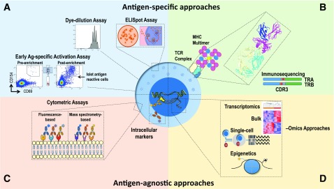
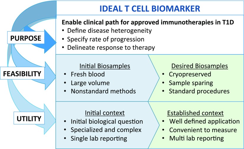
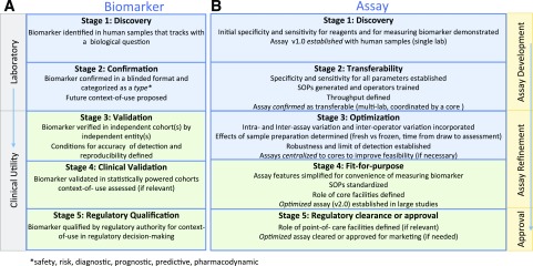
Type 1 diabetes (T1D) results from the progressive destruction of pancreatic β-cells in a process mediated primarily by T lymphocytes. The T1D research community has made dramatic progress in understanding the genetic basis of the disease as well as in the development of standardized autoantibody assays that inform both disease risk and progression. Despite these advances, there remains a paucity of robust and accepted biomarkers that can effectively inform on the activity of T cells during the natural history of the disease or in response to treatment. In this article, we discuss biomarker development and validation efforts for evaluation of T-cell responses in patients with and at risk for T1D as well as emerging technologies. It is expected that with systematic planning and execution of a well-conceived biomarker development pipeline, T-cell-related biomarkers would rapidly accelerate disease progression monitoring efforts and the evaluation of intervention therapies in T1D.


Figures

Figure 1
Figure 1
Methods for assessing T-cell biomarkers in T1D. Experimental approaches include assays for assessing both antigen-specific (A and B) and antigen-agnostic features of T cells (C and D). A: Assays for monitoring antigen-specific T-cell activation, proliferation, and cytokine production. B: HLA class I or II multimers loaded with autoantigenic peptides facilitate the detection, phenotyping, and downstream molecular analysis of antigen-specific T cells. Shown is a rendering of the 1E6 TCR recognizing a preproinsulin peptide in the HLA-A*0201 binding groove (85). Immunosequencing of the TRA and TRB genes encoding the V (blue), D-J (red/yellow and gray), and C (green) regions of the TCR-α and TCR-β chains, respectively, facilitates characterization of the TCR reactivity antigen-binding pocket, as determined from the highly polymorphic TRB complementarity-determining region 3 (CDR3; red/yellow) or by complete α/β-chain pairing. C: Flow cytometric approaches employing antibodies conjugated to fluorescent molecules or metals (via mass cytometry) can be used to phenotype a large array of surface and intracellular markers. D: Both bulk- and single-cell technologies facilitate phenotypic, transcriptional, and epigenetic profiling of T cells. Recent advances now facilitate integration of these methodologies at the single-cell resolution, providing high-parameter T-cell biomarkers with molecular resolution.
Figure 2
Figure 2
Process considerations for developing informative T-cell biomarkers. Biomarker development begins by defining the purpose of the biomarker and then assessing feasibility and utility. The boxes in blue (left) indicate features of candidate T-cell biomarkers and the assays used to detect them that are in development, whereas green boxes (right) highlight the key features required of validated T-cell biomarkers and associated assays. Feasibility includes considerations for sample-sparing assays utilizing cryopreserved biobanked samples.
Figure 3
Figure 3
Proposed stages of development for T1D biomarkers and assays (consensus view of the authors). Biomarkers and their associated assays have parallel and independent lines of development, ideally converging at the stage of biomarker validation using fit-for-purpose assays for reliable use by the scientific community. A: A biomarker must successfully pass through stage 3 to be considered validated for research purposes. If a biomarker is a candidate for regulatory decision-making, subsequent stages of development (stages 4–5) must be completed. B: All assays should ideally achieve fit-for-purpose status (stage 4) for widespread use to measure a validated biomarker. In specific instances, where an assay has achieved approval for marketing purposes, it must be cleared by regulatory bodies (stage 5). SOPs, standard operating procedures.

References

    1. McKinney EF, Lee JC, Jayne DR, Lyons PA, Smith KG. T-cell exhaustion, co-stimulation and clinical outcome in autoimmunity and infection. Nature 2015;523:612–616 - PMC - PubMed
    1. Roep BO, Peakman M. Antigen targets of type 1 diabetes autoimmunity. Cold Spring Harb Perspect Med 2012;2:a007781 - PMC - PubMed
    1. Unger WW, Velthuis J, Abreu JR, et al. . Discovery of low-affinity preproinsulin epitopes and detection of autoreactive CD8 T-cells using combinatorial MHC multimers. J Autoimmun 2011;37:151–159 - PubMed
    1. Yang J, Chow IT, Sosinowski T, et al. . Autoreactive T cells specific for insulin B:11-23 recognize a low-affinity peptide register in human subjects with autoimmune diabetes. Proc Natl Acad Sci U S A 2014;111:14840–14845 - PMC - PubMed
    1. Scotto M, Afonso G, Østerbye T, et al. . HLA-B7-restricted islet epitopes are differentially recognized in type 1 diabetic children and adults and form weak peptide-HLA complexes. Diabetes 2012;61:2546–2555 - PMC - PubMed

Publication types