. 2019 Aug 22;54(2):1802441.
doi: 10.1183/13993003.02441-2018. Print 2019 Aug.

Proliferating SPP1/MERTK-expressing macrophages in idiopathic pulmonary fibrosis

Affiliations

Proliferating SPP1/MERTK-expressing macrophages in idiopathic pulmonary fibrosis

Christina Morse et al. Eur Respir J. .

Abstract

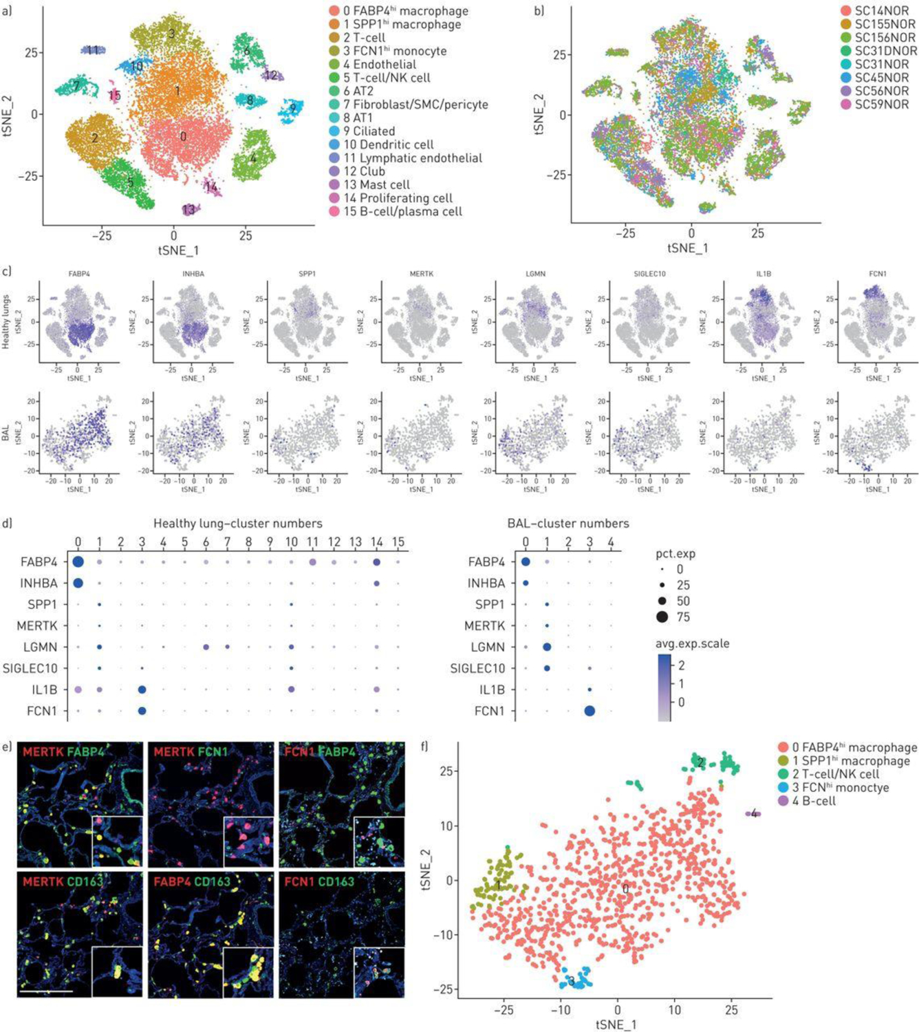
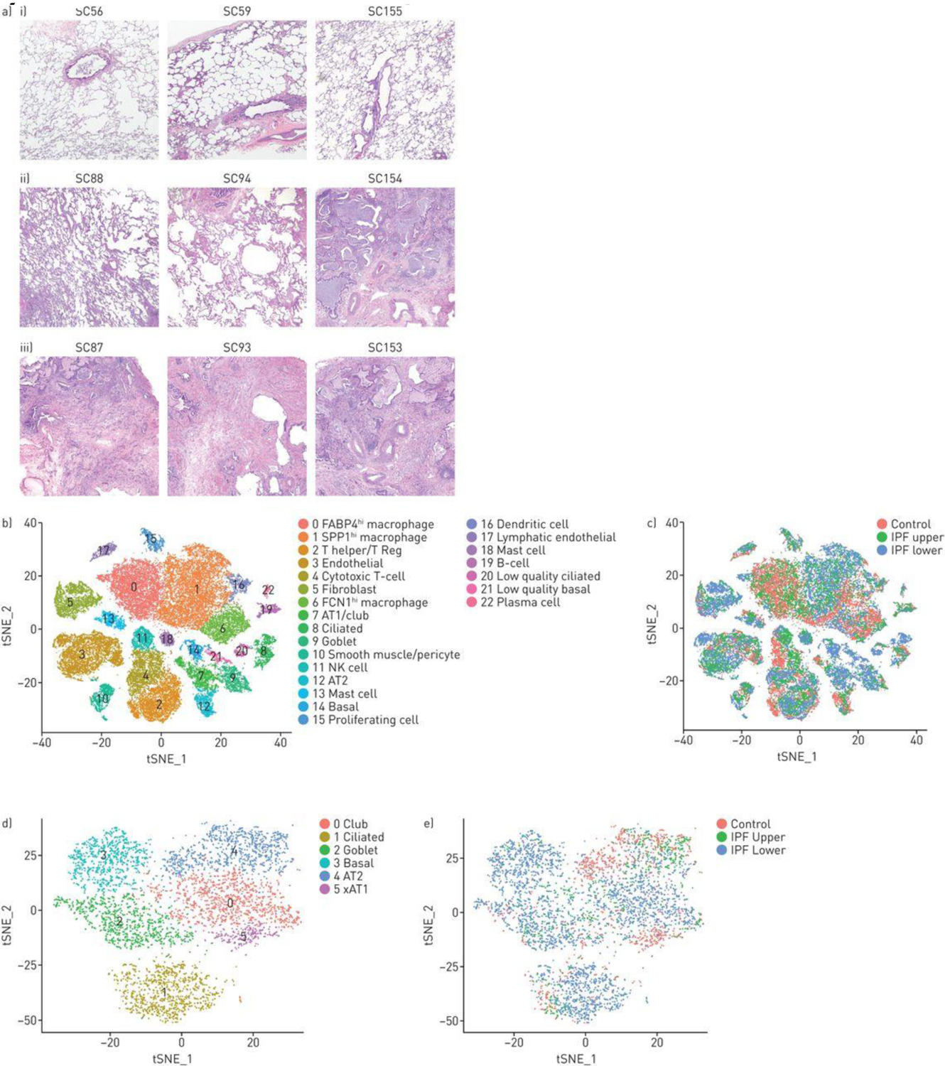
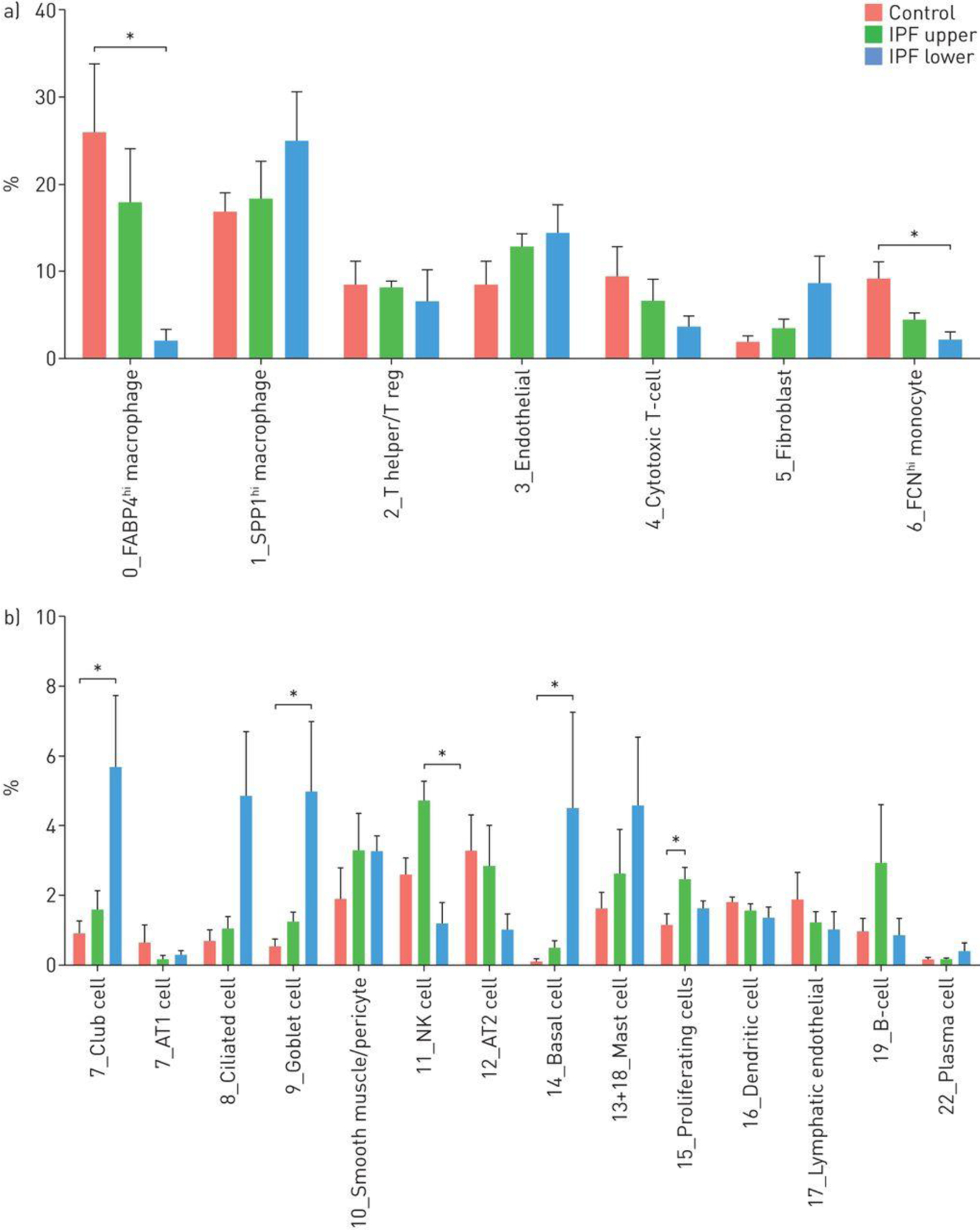
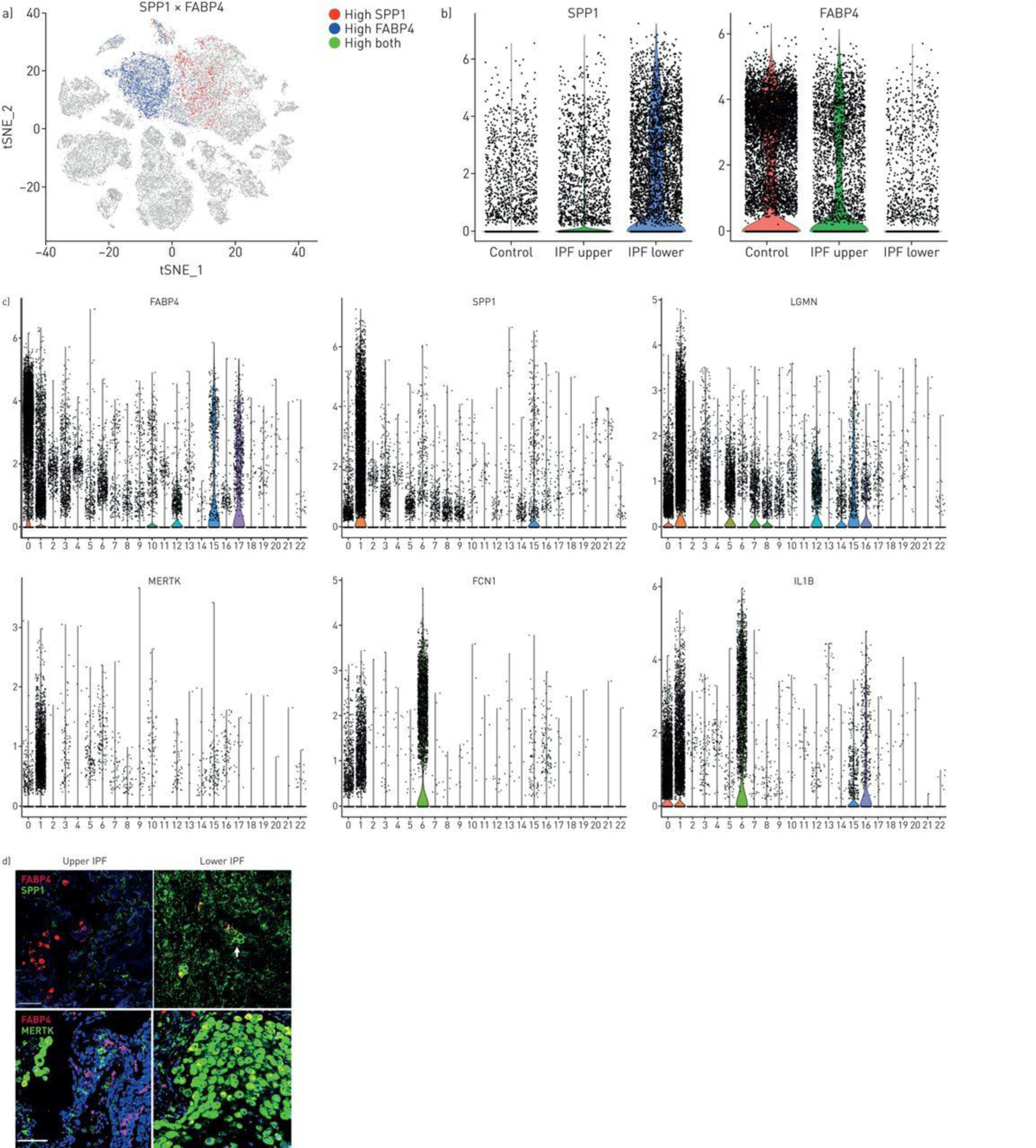
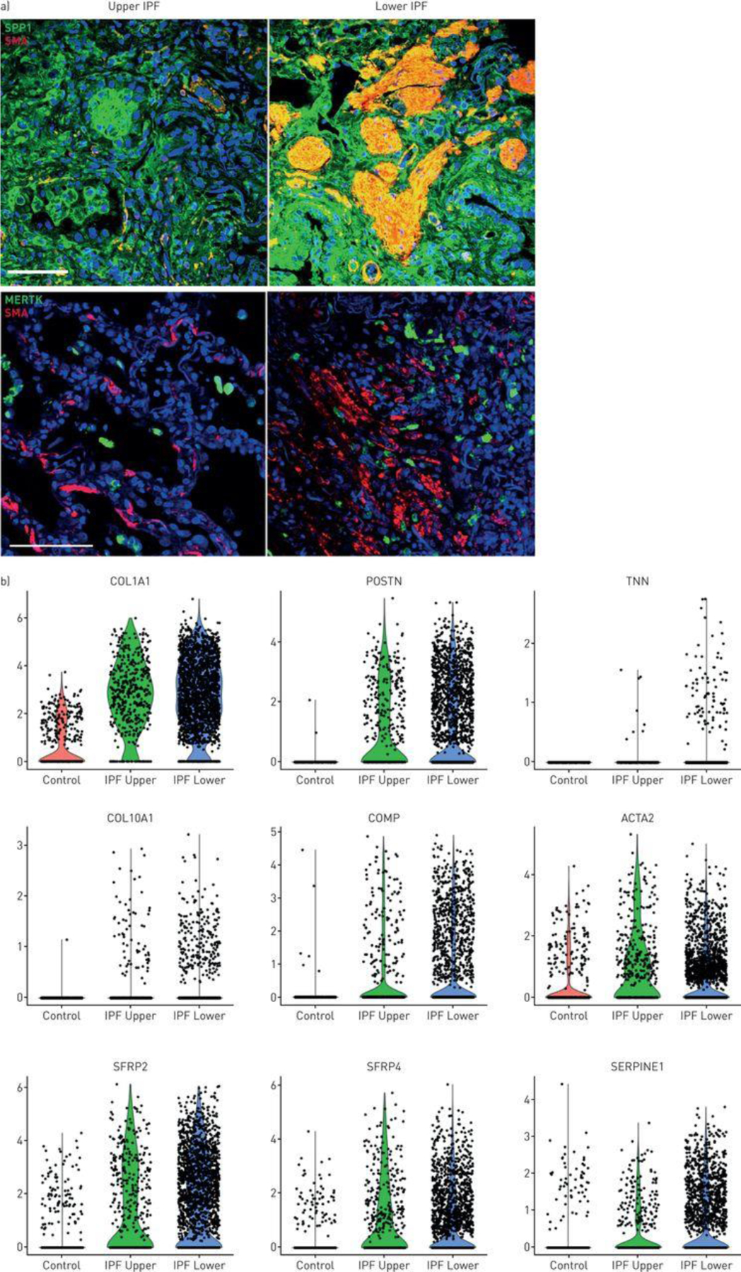
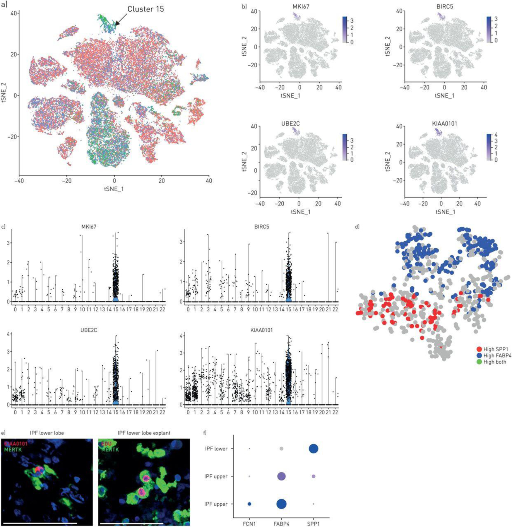
A comprehensive understanding of the changes in gene expression in cell types involved in idiopathic pulmonary fibrosis (IPF) will shed light on the mechanisms underlying the loss of alveolar epithelial cells and development of honeycomb cysts and fibroblastic foci. We sought to understand changes in IPF lung cell transcriptomes and gain insight into innate immune aspects of pathogenesis.We investigated IPF pathogenesis using single-cell RNA-sequencing of fresh lung explants, comparing human IPF fibrotic lower lobes reflecting late disease, upper lobes reflecting early disease and normal lungs.IPF lower lobes showed increased fibroblasts, and basal, ciliated, goblet and club cells, but decreased alveolar epithelial cells, and marked alterations in inflammatory cells. We found three discrete macrophage subpopulations in normal and fibrotic lungs, one expressing monocyte markers, one highly expressing FABP4 and INHBA (FABP4hi), and one highly expressing SPP1 and MERTK (SPP1hi). SPP1hi macrophages in fibrotic lower lobes showed highly upregulated SPP1 and MERTK expression. Low-level local proliferation of SPP1hi macrophages in normal lungs was strikingly increased in IPF lungs.Co-localisation and causal modelling supported the role for these highly proliferative SPP1hi macrophages in activation of IPF myofibroblasts in lung fibrosis. These data suggest that SPP1hi macrophages contribute importantly to lung fibrosis in IPF, and that therapeutic strategies targeting MERTK and macrophage proliferation may show promise for treatment of this disease.


Conflict of interest statement

Conflict of interest: C. Morse has nothing to disclose. Conflict of interest: T. Tabib has nothing to disclose. Conflict of interest: J. Sembrat has nothing to disclose. Conflict of interest: K. Buschur has nothing to disclose. Conflict of interest: H. Trejo Bittar has nothing to disclose. Conflict of interest: E. Valenzi has nothing to disclose. Conflict of interest: Y. Jiang has nothing to disclose. Conflict of interest: D.J. Kass reports grants from NIH, during the conduct of the study; grants from Regeneron, outside the submitted work. Conflict of interest: K. Gibson has nothing to disclose. Conflict of interest: W. Chen has nothing to disclose. Conflict of interest: A. Mora has nothing to disclose. Conflict of interest: P.V. Benos has nothing to disclose. Conflict of interest: M. Rojas has nothing to disclose. Conflict of interest: R. Lafyatis has nothing to disclose.

Figures

Figure 1.
Figure 1.
Characterisation of normal lung macrophages. T-distributed stochastic neighbour embedding (T-SNE) of normal lung cells indicates 10 cell types including a large central grouping of macrophage/monocytes/dendritic cells composed of clusters 0, 1, 3 and 10 (panel a) All four of these clusters express AIF1 and all but the dendritic cell cluster (cluster 10) express CD163 (supplementary figure S1). The subject of origin for each of the samples in indicated by a different colour in panel b. The three different macrophage/monocyte clusters (clusters 0, 1 and 3) can be distinguished in feature plots (panel c, purple intensity indicates level of gene expression) and dot plots (panel d, dot size indicates percent of cells expressing and the intensity of purple the level of expression) by expression of FABP4 and INHBA (FABP4hi macrophages, cluster 0); SPP1, MERTK, LGMN and SIGLEC10 (SPP1hi macrophages, cluster 1); or IL1B and FCN1 (FCN1hi monocyte/macrophages, cluster 3). Dendritic cells, expressing CD1C, are found in cluster 10 (see also supplementary figure S1). Macrophage monocyte cell types in bronchoalveolar lavage (BAL) clusters can be distinguished by gene expression (panels c and d), showing FABP4 and INHBA (FABP4hi macrophages, cluster 0); SPP1, MERTK, LGMN and SIGLEC10 (SPP1hi macrophages, cluster 1); or IL1B and FCN1 (FCN1hi monocyte/macrophages, cluster 3). Double immunoflorescent staining for MERTK, FABP4 and FCN1 encoded proteins in serial sections show mostly discrete staining for these three macrophage populations, all three co-stain for CD163. Scale bar=500 μm (panel e). BAL cells from a healthy subject clustered by t-SNE show discrete clusters of macrophages (panel f).
Figure 2.
Figure 2.
Combined t-distributed stochastic neighbour embedding (t-SNE) analysis of single-cell transcriptomes from three normal, three idiopathic pulmonary fibrosis (IPF) upper and three IPF lower lobes. a) Tissue sections of i) three control lung samples show preserved lung architecture with no significant pleural, subpleural or interstitial fibrosis and no increase in inflammatory cells. Tissue sections from patients with IPF showed iii) usual interstitial pneumonia lower lobes with a predominant cicatricial process and complete replacement of the lung architecture by scar tissue with honeycomb change, increased inflammatory cells and fibroblastic foci. ii) In comparison, the upper lobes showed relatively uninvolved lung tissue with preserved architecture and only mild interstitial organisation and mildly increased interstitial mononuclear cells suggestive of acute/subacute inflammation in samples SC88 and SC94 with more extensive fibrosis in SC154. Scale bar=1 mm. b) The t-SNE plot shows 20 clusters, with cell types identified by marker genes (supplementary figures S13 and S14). c,e) The tissue origin of the cells stratified by the type of tissue (control, IPF upper or IPF lower lobes) is indicated by different colouring of the cells. d) Reclustering of epithelial cells, showing the discrete clusters of club, ciliated (cluster 1) goblet and AT1 and AT2 cells, as identified by marker genes (supplementary figure S15).
Figure 3.
Figure 3.
Percentages of cell types captured in control, upper idiopathic pulmonary fibrosis (IPF) and lower IPF lung samples. The average proportion of cell subgroups as a percentage of total cells analysed, in control, upper IPF lobes and lower IPF lobes are shown for each cluster as shown in figure 2a and subclusters as shown in figure 2d. *: p<0.05.
Figure 4.
Figure 4.
Macrophage populations in idiopathic pulmonary fibrosis (IPF) lungs. a) SPP1 and FABP4 expression define two discrete populations in IPF and normal lungs. b) (and supplementary figure S18) SPP1hi macrophages express more SPP1 and make up a higher percentage of cells in lower lobes, while FABP4hi macrophages are a higher percentage of the cells in healthy and IPF upper lobes. c) Violin plots of combined IPF/control lung data show expression of FABP4, SPP1, LGMN, MERTK, FCN1 and IL1B limited mainly to macrophage populations (clusters 0, FABP4hi macrophages; cluster 1, SPP1hi macrophages and cluster 6, FCN1hi monocyte/macrophages; figure 2). d) (and supplementary figure S19) FABP4 expression is increased in FABP4hi macrophages, although it is also expressed at lower levels in SPP1hi macrophages; SPP1, MERTK and LGMN show highly increased expression in SPP1hi macrophages; FCN1 and IL1B are most highly expressed in FCN1hi monocyte/macrophages. Immunofluorescent staining for SPP1 shows macrophages embedded in the SPP1/osteopontin matrix; while staining for MERTK and FABP4 reveals these largely two discrete macrophage populations with increased FABP4-staining macrophages in upper lobes and increased MERTK macrophages in lower lobes. Scale bar=100 μm.
Figure 5.
Figure 5.
SPP1 macrophages and myofibroblasts in idiopathic pulmonary fibrosis (IPF) lungs. a) Fibroblastic foci in IPF lungs were stained with α-smooth muscle actin (SMA; red). Macrophages were stained with SPP1 or MERTK as indicated (green). b) Violin plots, indicating expression of several genes associated with fibrosis in control, IPF upper and IPF lower lungs.
Figure 6.
Figure 6.
Proliferating cells in idiopathic pulmonary fibrosis (IPF) and normal lungs. a) Proliferating cells from control and IPF lungs form a distinct cluster (cluster 15, green G2/M phase cells marked by arrow; see also figure 2b, cluster 15). b, c) Cluster 15 cells highly express MKI67 (Ki-67), BIRC5, UBE2C and KIAA0101. d) Reclustering of cluster 15 cells shows macrophages expressing either FABP4 or SPP1. Using KIAA as a marker for proliferating cells, both FABP4 and SPP1hi cells are proliferating in this cluster. e) MERTK-expressing macrophages from IPF lower lobe lung explant co-stained with MERTK and proliferation marker KIAA0101. Macrophages labelled in vitro by incubation with EdU (red), co-stained by immunofluorescent with MERTK (green). f) Examining gene expression by only the proliferating macrophage subset shows low level proliferation of FCN1/(IL1B-expressing) macrophages in control lungs; primarily proliferation of FCN1/(IL1B-expressing) macrophages and FABP4hi macrophages in IPF upper lobes and primarily proliferation of SPP1hi macrophages in IPF lower lobes (size of dot indicates the proportion of cells and intensity of purple the relative level of gene expression). Scale bar=100 μm.

Comment in

References

    1. Blackwell TS, Tager AM, Borok Z, Moore BB, Schwartz DA, Anstrom KJ, Bar-Joseph Z, Bitterman P, Blackburn MR, Bradford W, Brown KK, Chapman HA, Collard HR, Cosgrove GP, Deterding R, Doyle R, Flaherty KR, Garcia CK, Hagood JS, Henke CA, Herzog E, Hogaboam CM, Horowitz JC, King TE Jr., Loyd JE, Lawson WE, Marsh CB, Noble PW, Noth I, Sheppard D, Olsson J, Ortiz LA, O’Riordan TG, Oury TD, Raghu G, Roman J, Sime PJ, Sisson TH, Tschumperlin D, Violette SM, Weaver TE, Wells RG, White ES, Kaminski N, Martinez FJ, Wynn TA, Thannickal VJ, Eu JP. Future directions in idiopathic pulmonary fibrosis research. An NHLBI workshop report. American journal of respiratory and critical care medicine 2014: 189(2): 214–222. - PMC - PubMed
    1. Sisson TH, Mendez M, Choi K, Subbotina N, Courey A, Cunningham A, Dave A, Engelhardt JF, Liu X, White ES, Thannickal VJ, Moore BB, Christensen PJ, Simon RH. Targeted injury of type II alveolar epithelial cells induces pulmonary fibrosis. American journal of respiratory and critical care medicine 2010: 181(3): 254–263. - PMC - PubMed
    1. Zoz DF, Lawson WE, Blackwell TS. Idiopathic pulmonary fibrosis: a disorder of epithelial cell dysfunction. The American journal of the medical sciences 2011: 341(6): 435–438. - PMC - PubMed
    1. Evans CM, Fingerlin TE, Schwarz MI, Lynch D, Kurche J, Warg L, Yang IV, Schwartz DA. Idiopathic Pulmonary Fibrosis: A Genetic Disease That Involves Mucociliary Dysfunction of the Peripheral Airways. Physiological reviews 2016: 96(4): 1567–1591. - PMC - PubMed
    1. Rock JR, Gao X, Xue Y, Randell SH, Kong YY, Hogan BL. Notch-dependent differentiation of adult airway basal stem cells. Cell stem cell 2011: 8(6): 639–648. - PMC - PubMed

Publication types