

Mechanism of gut microbiota and Axl/SOCS3 in experimental autoimmune encephalomyelitis
- PMID: 31221818
- PMCID: PMC6603274
- DOI: 10.1042/BSR20190228
Mechanism of gut microbiota and Axl/SOCS3 in experimental autoimmune encephalomyelitis
Abstract
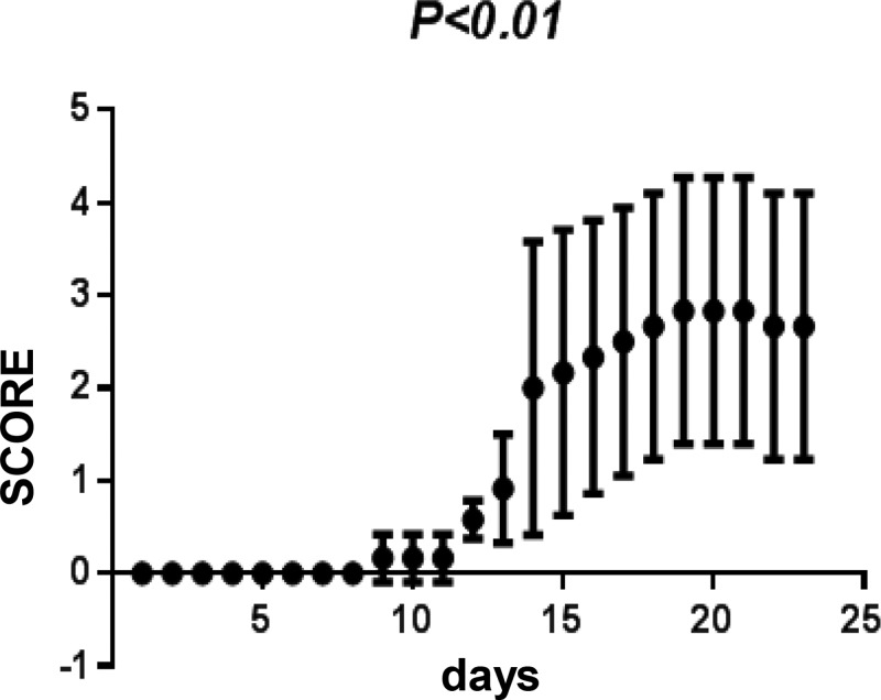
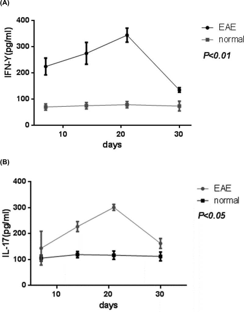
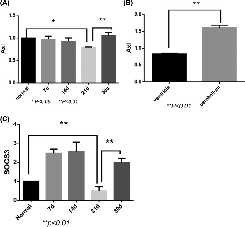
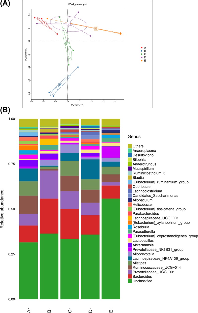
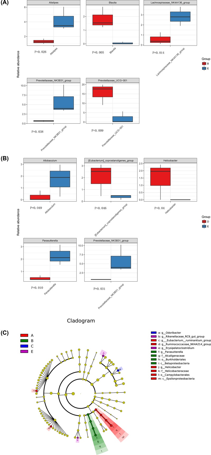
Multiple sclerosis (MS) is an immune-mediated disease of the central nervous system (CNS). The present study explored the role of intestinal microbiota in the initiation and propagation of mice induced by experimental autoimmune encephalomyelitis (EAE), an animal model of multiple sclerosis. 48 C57BL/6 were randomly divided into control group and EAE group. The changes of body weight and the scores of neurological function were recorded. The mRNA expression of the receptor tyrosine kinase subfamily (AXL) was detected by real-time quantitative PCR. The levels of IL-17 and IFN-γ in blood samples were examined by ELISA. The intestinal microbial composition of mice at different time points during the EAE induction was analyzed by 16S rRNA gene-based sequencing. In EAE group, the body weight began to reduce at day 3 and neurological symptoms began to appear at day 7 after EAE induction. The levels of IL-17 and IFN-γ in EAE group reached the peak at day 21 and then decreased gradually. However, the expression of Axl and SOCS3 reached the lowest level at day 21 and then increased gradually. The microbiome analyses revealed that the abundances of Alistipes, Blautia, and Lachnospiraceae_NK4A136_group were significantly changed at day 14, whereas the abundances of Allobaculum, Eubacterium and Helicobacter were significantly changed at day 30 of EAE induction. The prevotellaceae_NK3B31_group may be key bacteria that contribute to the development of MS. Regulation of intestinal microbiota composition can become a new therapeutic target for the treatment of MS.
Keywords: Gut microbiota; Inflammatory inhibitors; Receptor tyrosine kinase subfamily; experimental autoimmune encephalomyelitis.
© 2019 The Author(s).
Conflict of interest statement
The authors declare that there are no competing interests associated with the manuscript.
Figures
Similar articles
-
Loss of the receptor tyrosine kinase Axl leads to enhanced inflammation in the CNS and delayed removal of myelin debris during experimental autoimmune encephalomyelitis.J Neuroinflammation. 2011 May 15;8:49. doi: 10.1186/1742-2094-8-49. J Neuroinflammation. 2011. PMID: 21569627 Free PMC article.
-
Lentivirus-mediated estrogen receptor α overexpression in the central nervous system ameliorates experimental autoimmune encephalomyelitis in mice.Int J Mol Med. 2013 May;31(5):1209-21. doi: 10.3892/ijmm.2013.1306. Epub 2013 Mar 15. Int J Mol Med. 2013. PMID: 23525227
-
Novel anti-inflammatory compounds that alleviate experimental autoimmune encephalomyelitis.Phytomedicine. 2025 Apr;139:156544. doi: 10.1016/j.phymed.2025.156544. Epub 2025 Feb 28. Phytomedicine. 2025. PMID: 40023067
-
Gut Microbiota in Multiple Sclerosis and Experimental Autoimmune Encephalomyelitis: Current Applications and Future Perspectives.Mediators Inflamm. 2018 Apr 2;2018:8168717. doi: 10.1155/2018/8168717. eCollection 2018. Mediators Inflamm. 2018. PMID: 29805314 Free PMC article. Review.
-
The second brain: The connection between gut microbiota composition and multiple sclerosis.J Neuroimmunol. 2021 Nov 15;360:577700. doi: 10.1016/j.jneuroim.2021.577700. Epub 2021 Aug 24. J Neuroimmunol. 2021. PMID: 34482269 Review.
Cited by
-
Intestinal Flora and Disease Mutually Shape the Regional Immune System in the Intestinal Tract.Front Immunol. 2020 Apr 3;11:575. doi: 10.3389/fimmu.2020.00575. eCollection 2020. Front Immunol. 2020. PMID: 32318067 Free PMC article. Review.
-
Integrating fecal metabolomics and intestinal microbiota to study the mechanism of cannabidiol in the treatment of idiopathic pulmonary fibrosis.Front Pharmacol. 2024 Feb 6;15:1358626. doi: 10.3389/fphar.2024.1358626. eCollection 2024. Front Pharmacol. 2024. PMID: 38379898 Free PMC article.
-
Ursolic Acid Ameliorates Spinal Cord Injury in Mice by Regulating Gut Microbiota and Metabolic Changes.Front Cell Neurosci. 2022 May 4;16:872935. doi: 10.3389/fncel.2022.872935. eCollection 2022. Front Cell Neurosci. 2022. PMID: 35602557 Free PMC article.
-
HLA Class II Polymorphisms Modulate Gut Microbiota and Experimental Autoimmune Encephalomyelitis Phenotype.Immunohorizons. 2021 Aug 11;5(8):627-646. doi: 10.4049/immunohorizons.2100024. Immunohorizons. 2021. PMID: 34380664 Free PMC article.
-
The Promotion of Humoral Immune Responses in Humans via SOCS1-Mediated Th2-Bias Following SARS-CoV-2 Vaccination.Vaccines (Basel). 2023 Nov 20;11(11):1730. doi: 10.3390/vaccines11111730. Vaccines (Basel). 2023. PMID: 38006062 Free PMC article.
References
MeSH terms
Substances
LinkOut - more resources
Full Text Sources
Medical
Research Materials
Miscellaneous