. 2019 Jun 21;364(6446):1156-1162.
doi: 10.1126/science.aaw3145.

The glycan CA19-9 promotes pancreatitis and pancreatic cancer in mice

Affiliations

The glycan CA19-9 promotes pancreatitis and pancreatic cancer in mice

Dannielle D Engle et al. Science. .

Abstract

Glycosylation alterations are indicative of tissue inflammation and neoplasia, but whether these alterations contribute to disease pathogenesis is largely unknown. To study the role of glycan changes in pancreatic disease, we inducibly expressed human fucosyltransferase 3 and β1,3-galactosyltransferase 5 in mice, reconstituting the glycan sialyl-Lewisa, also known as carbohydrate antigen 19-9 (CA19-9). Notably, CA19-9 expression in mice resulted in rapid and severe pancreatitis with hyperactivation of epidermal growth factor receptor (EGFR) signaling. Mechanistically, CA19-9 modification of the matricellular protein fibulin-3 increased its interaction with EGFR, and blockade of fibulin-3, EGFR ligands, or CA19-9 prevented EGFR hyperactivation in organoids. CA19-9-mediated pancreatitis was reversible and could be suppressed with CA19-9 antibodies. CA19-9 also cooperated with the KrasG12D oncogene to produce aggressive pancreatic cancer. These findings implicate CA19-9 in the etiology of pancreatitis and pancreatic cancer and nominate CA19-9 as a therapeutic target.


Conflict of interest statement

Competing Interests. CSHL owns a pending US provisional patent application directed to the use of CA19–9 antibodies to treat pancreatitis, which was filed on behalf of CSHL under a license from CSHL to MabVax Therapeutics Holdings, Inc. DAT serves on the Scientific Advisory Board of Leap Therapeutics, Surface Oncology, and Bethyl Laboratory, which is not related to the subject matter of this manuscript. DAT also serves on the Board of Scientific Advisors for the NCI, the Scientific Advisory Board of AACR, the Scientific Advisory Council of Stand Up to Cancer, and the Scientific Advisory Committee of the Georg-Speyer-Haus Institute for Tumor Biology and Experimental Therapy. DAT is a distinguished scholar of the Lustgarten Foundation and Director of the Lustgarten Foundation-designated Laboratory of Pancreatic Cancer Research.

Figures

Fig. 1.
Fig. 1.. FUT3 with β3GALT5 expression enables CA19–9 production in engineered mouse pancreatic cancer cells.
(A) Ectopic FUT3 induces Lewisx (Lex) but not CA19–9/sialyl-Lewisa (sLea) expression in mouse PDAC cells by flow cytometric analysis. The Lex and CA19–9/sLea positive human cell line Colo205 and Lex and CA19–9/sLea negative parental KPC cell lines are shown. (B) CA19–9 flow cytometry of mouse PDAC cells stably and constitutively expressing FUT3 with β3GALT5 (“FB”) compared to the isotype control antibody. (C) Overlap between CA19–9 protein carriers identified in 3 out of 3 human PDAC cell lines (n = 926) with three independent mouse PDAC cell lines expressing FUT3 and β3GALT5.
Fig. 2.
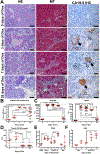
Fig. 2.. CA19–9 expression promotes pancreatitis in mice.
(A) Histologic evaluation of C;RLSL;F mice by hematoxylin and eosin staining (H&E), Masson’s trichrome staining (MT, blue indicates collagen deposition) and CA19–9 expression (open arrow, CA19–9+ duct; closed arrow, CA19–9+ islet) by IHC following treatment with Dox. Scale bars = 50μm. (B) Quantification of the percentage pancreatic area exhibiting histologic signs of pancreatitis following treatment of C;RLSL;F mice with Dox. (C) Circulating levels (U/L) of the pancreatic enzymes amylase and lipase in C;RLSL;F mice following treatment with Dox (Days). The dotted line indicates the threshold elevation required for the diagnosis of pancreatitis. The dashed line indicates the maximum level of detection possible for amylase. (D) The circulating level of CA19–9 (U/ml) following treatment of C;RLSL;F mice with Dox. Values that exceed 37U/ml (dotted line) are elevated. (E) Immune cell infiltration evaluated by flow cytometry in mice treated with PBS or Cerulein for 2 days followed by a 1 or 3 day recovery period (C57Bl/6j, n = 5, 5, and 5, respectively) and C;RLSL;F mice treated with Dox (n = 4, 4, and 6, respectively) compared to “genetically negative” controls (GN, n = 3 and 3, respectively. Outlier analysis using Grubb’s method identified one data point (triangle symbol, GN, 3 days of Dox) as an outlier. (F) EdU incorporation in the pancreas was evaluated by flow cytometry in C;RLSL;F mice (n = 3, 3, and 4, respectively) and genetically negative littermate controls (GN, n = 3 and 2, respectively) following treatment with Dox. Middle horizontal red lines represent the mean and error bars represent the standard deviation; each data point represents a measurement from an individual mouse. * p<0.05, ** p<0.01, *** p<0.001, **** p<0.0001 for multiple comparisons using Holm-Sidak’s procedure following a one-way ANOVA.
Fig. 3.
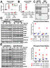
Fig. 3.. CA19–9 expression activates EGFR signaling.
(A) Representative phosphorylated-EGFR IHC in C;RLSL;F mice that were treated for 0 (n = 4) or 3 (n = 5) days with Dox. Insets are higher magnification of pancreatic ducts. Scale bars = 50μm. (B) GSEA of C;RLSL;F organoids identified enrichment in PI3K/AKT/MTOR and ECM Receptor signaling and downstream effector pathways. (C) C;RLSL;F organoids (n = 3 biological replicates) and genetically negative organoids (GN, n = 2 biological replicates) were evaluated by immunoblot for the activation of signaling pathways following treatment with Dox. Blots are representative of three technical replicates, three biological replicates of C;RLSL;F organoids and two biological replicates of GN organoids. (D) Quantification of changes to the ratio of phosphorylated to total protein is shown for Figures 3E, 3F, and 3G. (E-F) Organoids from GN littermates were incubated with conditioned media from C;RLSL;F organoids previously treated with Dox (24 hours) in the presence of Fc-control or Egfr-conjugated Fc and evaluated by immunoblot. (G) C;RLSL;F organoids (n = 3 biological replicates) were treated with Dox (Hours) in the presence of isotype control (MOPC21) or CA19–9 blocking mAb (NS19–9) and evaluated by immunoblot. Blots are representative of two technical and three biological replicates.
Figure 4.
Figure 4.. CA19–9 modified Fibulin 3 activates Egfr.
(A) IP/MS of EGFR complexes from C;RLSL;F (n=3) and GN organoid (n=2) whole cell lysates following Dox administration. (B) CA19–9 protein carrier IP/MS analysis of Fibulin 3 from conditioned media from C;RLSL;F organoids treated with Dox for 0 – 8 hours (n = 3) relative to untreated and treated GN organoids (n = 1) using 5B1 CA19–9 antibody clone. (C) Immunblot of immunoprecipitated Fibulin 3 (Flag) following incubation with lysates from pancreatic cancer cells lacking Flag-Fbln3 expression. (D) C;RLSL;F organoids (n = 3) treated with Dox in the presence of MOPC21 or three independent Fibulin 3 antibodies and evaluated by immunoblot. Changes to the ratio of phosphorylated to total protein levels are included. (E) Organoids transduced with hairpins to GFP or to Fbln3 were immunoblotted following Dox treatment. Changes to the ratio of phosphorylated to total protein levels are included. Middle horizontal red lines represent the mean and error bars represent the standard deviation; each data point represents a biological replicate. * p<0.05 by unpaired, two-tailed t test.
Figure 5.
Figure 5.. CA19–9 as a therapeutic target for the treatment of pancreatitis.
(A) H&E staining of representative areas from C;RLSL;F mice following treatment with human Isotype control (hIgG1, n = 5) or the CA19–9 antibody clone 5B1 (n = 5) for 8 days and Dox for the last 7 days. Scale bars = 100μm. (B) Serum amylase and lipase levels, and in C;RLSL;F mice treated with 5B1 or isotype control. (C) treated with either Isotype or 5B1 as described in Fig. 5A. Scale bars = 50μm. Middle horizontal red lines represent the mean and error bars represent the standard deviation; each data point represents a measurement from an individual mouse. p value determined using unpaired, parametric, two-tailed t test. * p<0.05, ** p<0.01, *** p<0.001, **** p<0.0001.
Figure 6.
Figure 6.. CA19–9 promotes rapid and aggressive pancreatic tumorigenesis.
(A) Survival curve for untreated (8 deaths out of 19 mice) and Dox treated K;C;RLSL:F mice (14 deaths out of 19 mice) and KC genetic negative controls (13 deaths out of 34 mice). p value determined by Log-rank Mantel-Cox test. (B) Representative histology of pancreatic tumors and metastatic lesions from Dox treated K;C;RLSL:F mice. Scale bars = 200μm. (C) Representative histology (scale bars = 100μm). (D) CA19–9 IHC (scale bars = 100μm), and (E) phospho-EGFR IHC (scale bars = 50μm) of the pancreata from K;C andK;C;RLSL:F mice following 2 – 4 weeks of Dox treatment.

Comment in

References

    1. Yadav D, Lowenfels AB, The epidemiology of pancreatitis and pancreatic cancer. Gastroenterology 144, 1252–1261 (2013). - PMC - PubMed
    1. Forsmark CE, Vege SS, Wilcox CM, Acute Pancreatitis. N Engl J Med 375, 1972–1981 (2016). - PubMed
    1. Frossard J-L, Steer ML, Pastor CM, Acute pancreatitis. The Lancet 371, 143–152 (2008). - PubMed
    1. van Brummelen SE, Venneman NG, van Erpecum KJ, VanBerge-Henegouwen GP, Acute idiopathic pancreatitis: does it really exist or is it a myth? Scandinavian journal of gastroenterology. Supplement, 117–122 (2003). - PubMed
    1. Lowenfels AB et al., Pancreatitis and the risk of pancreatic cancer. International Pancreatitis Study Group. N Engl J Med 328, 1433–1437 (1993). - PubMed

Publication types

MeSH terms

Substances