Review
. 2019 Jul;15(7):419-427.
doi: 10.1038/s41582-019-0222-0. Epub 2019 Jun 20.

SILK studies - capturing the turnover of proteins linked to neurodegenerative diseases

Affiliations
Review

SILK studies - capturing the turnover of proteins linked to neurodegenerative diseases

Ross W Paterson et al. Nat Rev Neurol. 2019 Jul.

Abstract

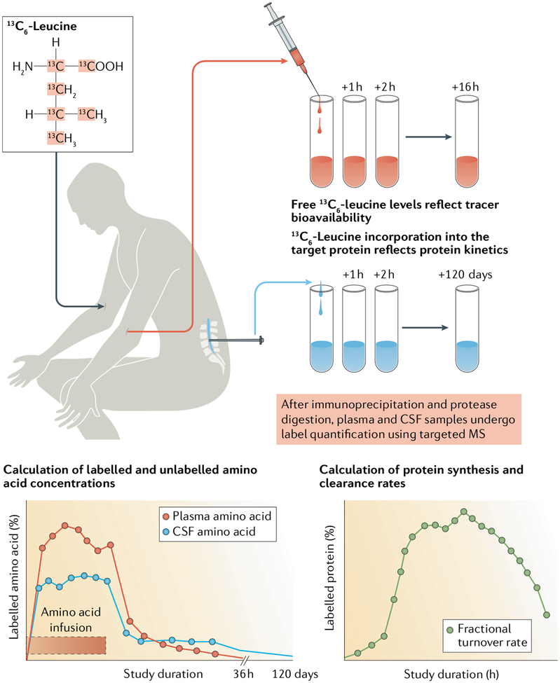
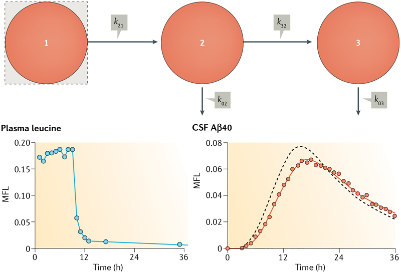
Alzheimer disease (AD) is one of several neurodegenerative diseases characterized by dysregulation, misfolding and accumulation of specific proteins in the CNS. The stable isotope labelling kinetics (SILK) technique is based on generating amino acids labelled with naturally occurring stable (that is, nonradioactive) isotopes of carbon and/or nitrogen. These labelled amino acids can then be incorporated into proteins, enabling rates of protein production and clearance to be determined in vivo and in vitro without the use of radioactive or chemical labels. Over the past decade, SILK studies have been used to determine the turnover of key pathogenic proteins amyloid-β (Aβ), tau and superoxide dismutase 1 (SOD1) in the cerebrospinal fluid of healthy individuals, patients with AD and those with other neurodegenerative diseases. These studies led to the identification of several factors that alter the production and/or clearance of these proteins, including age, sleep and disease-causing genetic mutations. SILK studies have also been used to measure Aβ turnover in blood and within brain tissue. SILK studies offer the potential to elucidate the mechanisms underlying various neurodegenerative disease mechanisms, including neuroinflammation and synaptic dysfunction, and to demonstrate target engagement of novel disease-modifying therapies.


Conflict of interest statement

Competing interests

H.Z. declares that he has served on scientific advisory boards for Roche Diagnostics, Samumed, CogRx and Wave and is one of the founders of Brain Biomarker Solutions in Gothenburg, which is funded by GU Ventures (a Swedish government-owned company managed by the University of Gothenburg); these activities are all unrelated to this article. R.J.B. declares that he, along with Washington University, has an equity ownership interest in C2N Diagnostics (a mass-spectrometry-based biotechnology company that holds patents on the stable isotope labelling kinetics (SILK) technique in the United States and other countries) and receives royalties related to SILK and blood plasma assay technologies licensed by Washington University to C2N Diagnostics. R.J.B. declares that he receives income from C2N Diagnostics for serving on its scientific advisory board. B.W.P. declares that he receives consultancy fees from C2N Diagnostics. T.M. and R.J.B. have licensed superoxide dismutase 1 SILK to C2N Diagnostics. N.C.W. holds a patent for SILK studies utilizing nanoscale secondary ion mass spectroscopy. K.Y. and T.W. declare that they are employed by C2N Diagnostics. The other authors declare no competing interests.

Figures

Fig. 1 |
Fig. 1 |. Stable isotope labelling kinetics methodology.
Stable isotope labelling kinetics (SILK) studies in humans involve infusion of an amino acid labelled with a stable isotope, such as 13C-leucine. To measure the turnover rate of a specific protein of interest, blood and cerebrospinal fluid (CSF) samples are collected at baseline and at appropriate intervals thereafter. Sampling intervals and duration are determined by the protein turnover rate. 13C-Leucine levels in blood reflect tracer bioavailability, whereas 13C-leucine levels in CSF and/or other body fluids reflect tracer incorporation into the target protein. To determine target protein kinetics, CSF samples usually undergo immunoprecipitation (to achieve target protein enrichment) and protease digestion, followed by label quantification using targeted mass spectroscopy (MS). These data are used to calculate the concentrations of labelled and unlabelled protein and, in compartmental modelling studies, to determine protein turnover rates.
Fig. 2 |
Fig. 2 |. Compartmental modelling of 13C-leucine kinetics in humans.
In this hypothetical model, compartment 1 represents the 13C-leucine tracer infusion site (typically blood) and compartment 2 represents the site of tracer incorporation into intermediates, such as amyloid precursor protein (APP), as well as transport delay processes. Compartment 3 represents the site of tracer incorporation into the protein of interest, such as amyloid-β (Aβ)40 in cerebrospinal fluid (CSF). The formula kxy is used to calculate the fraction of compartment y converted to compartment x per unit time, where 0 represents loss from the system. This calculation transforms the plasma 13C-leucine curve (left) into the Aβ40 13C-leucine curve (right). Mole fraction labelled (MFL) is a ‘forcing function’ that uses linear interpolation between measured time points to define tracer bioavailability for incorporation into target proteins. The compartmental model typically identifies a fractional turnover rate for the whole system, namely, the fraction of a rate-limiting compartment that is irreversibly removed per unit time. Mass flux rates along a pathway are calculated as the compartment mass multiplied by the rate constant. Specifically, if compartment 3 represents CSF Aβ, then the flux of Aβ through CSF (that is, the rate of appearance of Aβ into and disappearance of Aβ from CSF at steady state) is the product of the CSF concentration and k03. Compartmental modelling of stable isotope labelling kinetics (SILK) data consists of constructing a physiologically plausible model with optimized rate constants that provides an excellent fit to the experimental Aβ40 SILK data (solid line). In the CSF Aβ40 SILK data shown, gross systematic errors in the fit would result if k03 is increased by 20% (dashed line).
Fig. 3 |
Fig. 3 |. Data from stable isotope labelling kinetics studies of amyloid-β in humans.
Turnover of amyloid-β (Aβ)42 is accelerated in individuals with amyloidosis. The accumulation of 13C-leucine in Aβ peptides, termed the mole fraction labelled (MFL), was normalized to the plasma 13C-leucine plateau MFL for each person and averaged for 46 individuals without amyloidosis (age ± 1 s.d. 72.8 ± 6.3 years; part a) and 54 matched individuals with amyloidosis (age ± 1 s.d. 73.6 ± 6.9 years; part b). Mean ± 95% CI error bars are shown for Aβ40 (green) and Aβ42 (red). Aβ42 shows identical kinetics to Aβ40 in amyloidosis-negative individuals but shows faster turnover (that is, an earlier peak) than Aβ40 in amyloidosis-positive individuals. Aβ turnover slows with ageing. Normalized MFLs for Aβ40 (part c) and Aβ42 (part d) were averaged for amyloidosis-negative individuals in three different age groups: <60 years (blue, n = 23), 60–70 years (red, n = 27) and >70 years (green, n = 28). Turnover of Aβ peptides declines with increasing age, as shown by successively later, broader and lower peaks. Similar age-related trends were found in amyloidosis-positive individuals (not shown). Data points in all four graphs were fitted to comprehensive compartmental models describing the time course of 13C-leucine Aβ kinetics (solid lines). Adapted with permission from REF, Wiley-VCH.
Fig. 4 |
Fig. 4 |. Imaging scans showing incorporation of a stable isotope tracer into living brain tissue.
Stable isotope labelling kinetics (SILK)-secondary ion mass spectroscopy (SIMS) studies enable quantitative imaging of stable isotopes such as 13C at high spatial resolution (50–100 nm or <1 (μm3, much smaller than a single cell). The technique is capable of mass resolution between 13C and 12CH (13.00159 Da versus 13.00605 Da) or 13C14N and 12C15N (27.00276 Da versus 26.99644 Da). SILK-SIMS is also a high-sensitivity technique; <1% enrichment can be detected. By contrast, PET imaging obtains only average measurements at ~1 cm resolution, and the resolution of confocal microscopy is ~0.5–1.0 μm. The result is a nanometre-level histological map of in situ incorporation of a stable isotope tracer. a | Optical image of neurons stained with toluidine blue (magnification × 40). b | 12C14N ion map of the same field of view shown in panel a. Imaging carbon as cyanide ions (CN) results in improved image quality and contrast owing to the increased count rate per pixel attributable to its ionization. In addition, measuring carbon as CN produces an enhanced image of biological material owing to the abundance of carbon, nitrogen and C–N bonds, which almost entirely removes the carbon-rich and nitrogen-poor signals contributed by the embedding media. c | A false-colour 13C14N and 12C14N ion map showing the distribution of 13C in the sample. The natural abundance of 13C is 1.1%.

References

    1. Wilkinson DJ Historical and contemporary stable isotope tracer approaches to studying mammalian protein metabolism. Mass Spectrom. Rev 37, 57–80 (2018). - PMC - PubMed
    1. Gregg CT et al. Substantial replacement of mammalian body carbon with carbon-13. Life Sci 13, 775–782 (1973). - PubMed
    1. Haass C & Selkoe DJ Cellular processing of beta-amyloid precursor protein and the genesis of amyloid beta-peptide. Cell 75, 1039–1042 (1993). - PubMed
    1. Xie LL et al. Sleep drives metabolite clearance from the adult brain. Science 342, 373–377 (2013). - PMC - PubMed
    1. Roberts KF et al. Amyloid-β efflux from the central nervous system into the plasma. Ann. Neurol 76, 837–844 (2014). - PMC - PubMed

Publication types