

From basic perception deficits to facial affect recognition impairments in schizophrenia
- PMID: 31222063
- PMCID: PMC6586813
- DOI: 10.1038/s41598-019-45231-x
From basic perception deficits to facial affect recognition impairments in schizophrenia
Abstract
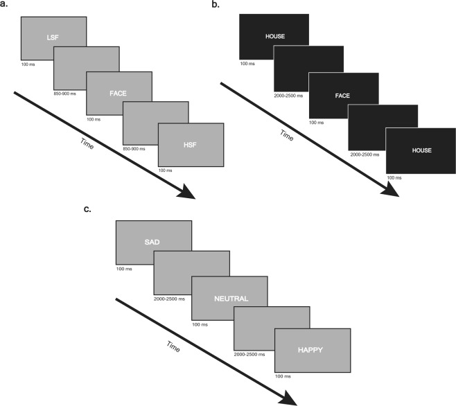
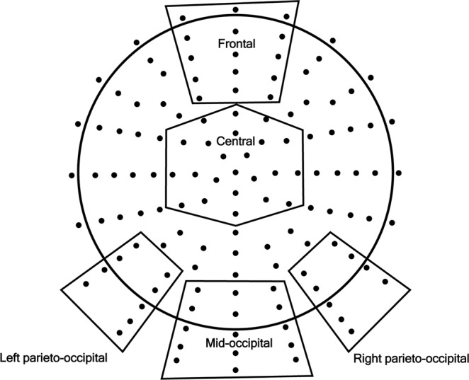
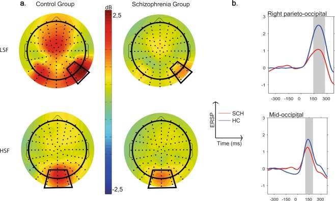
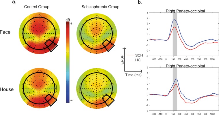
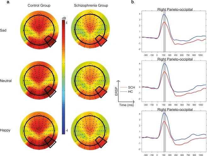
While impaired facial emotion recognition and magnocellular deficits in visual perception are core features of schizophrenia, their relationship is still unclear. Our aim was to analyze the oscillatory background of these processes and to investigate the connection between the magnocellular pathway deficit and the abnormal facial affect processing. Thirty-nine subjects with schizophrenia and forty socially matched healthy controls subjects were enrolled. A 128 channel EEG was recorded in three experimental tasks: first, participants viewed magnocellular biased low-spatial frequency (LSF) and parvocellular biased high-spatial frequency (HSF) Gabor-patches, then faces and houses were presented and in the third task a facial affect recognition task was presented with happy, sad and neutral faces. Event-related theta (4-7 Hz) synchronization (ERS) (i.e. an increase in theta power) by magnocellular biased stimuli was decreased in patients relative to controls, while no similar differences were found between groups in the parvocellular biased condition. ERS was significantly lower in patients compared to healthy controls both in the face and in the emotion recognition task. Theta ERS to magnocellular biased stimuli, but not to parvocellular biased stimuli, were correlated with emotion recognition performance. These findings indicate a bottom up disruption of face perception and emotion recognition in schizophrenia.
Conflict of interest statement
The authors declare no competing interests.
Figures
Similar articles
-
Probing the magnocellular and parvocellular visual pathways in facial emotion perception in schizophrenia.Psychiatry Res. 2017 Jul;253:38-42. doi: 10.1016/j.psychres.2017.03.031. Epub 2017 Mar 20. Psychiatry Res. 2017. PMID: 28342330
-
Early visual processing deficits in patients with schizophrenia during spatial frequency-dependent facial affect processing.Schizophr Res. 2015 Feb;161(2-3):314-21. doi: 10.1016/j.schres.2014.12.020. Epub 2014 Dec 29. Schizophr Res. 2015. PMID: 25553978
-
Event-related theta synchronization predicts deficit in facial affect recognition in schizophrenia.J Abnorm Psychol. 2014 Feb;123(1):178-89. doi: 10.1037/a0035793. J Abnorm Psychol. 2014. PMID: 24661169
-
[Neural mechanisms of face recognition: an event-related potential study].Brain Nerve. 2012 Jul;64(7):717-26. Brain Nerve. 2012. PMID: 22764343 Review. Japanese.
-
Face recognition in schizophrenia disorder: A comprehensive review of behavioral, neuroimaging and neurophysiological studies.Neurosci Biobehav Rev. 2015 Jun;53:79-107. doi: 10.1016/j.neubiorev.2015.03.006. Epub 2015 Mar 21. Neurosci Biobehav Rev. 2015. PMID: 25800172 Review.
Cited by
-
Disruption of early visual processing in amyloid-positive healthy individuals and mild cognitive impairment.Alzheimers Res Ther. 2023 Feb 28;15(1):42. doi: 10.1186/s13195-023-01189-7. Alzheimers Res Ther. 2023. PMID: 36855162 Free PMC article.
-
Evidence From Imaging Resilience Genetics for a Protective Mechanism Against Schizophrenia in the Ventral Visual Pathway.Schizophr Bull. 2022 May 7;48(3):551-562. doi: 10.1093/schbul/sbab151. Schizophr Bull. 2022. PMID: 35137221 Free PMC article.
-
An Event-Related Potential Investigation of Early Visual Processing Deficits During Face Perception in Youth at Clinical High Risk for Psychosis.Schizophr Bull. 2022 Jan 21;48(1):90-99. doi: 10.1093/schbul/sbab068. Schizophr Bull. 2022. PMID: 34111294 Free PMC article.
-
Weaker beta desynchronization indicates impaired emotion recognition in schizophrenia.Schizophrenia (Heidelb). 2025 Mar 7;11(1):39. doi: 10.1038/s41537-025-00591-4. Schizophrenia (Heidelb). 2025. PMID: 40055340 Free PMC article.
-
The 4D Space-Time Dimensions of Facial Perception.Front Psychol. 2020 Jul 28;11:1842. doi: 10.3389/fpsyg.2020.01842. eCollection 2020. Front Psychol. 2020. PMID: 32849084 Free PMC article. Review.
References
Publication types
MeSH terms
LinkOut - more resources
Full Text Sources
Medical
Molecular Biology Databases
