

Diphtheria Toxin A-Resistant Cell Lines Enable Robust Production and Evaluation of DTA-Encoding Lentiviruses
- PMID: 31222087
- PMCID: PMC6586843
- DOI: 10.1038/s41598-019-45481-9
Diphtheria Toxin A-Resistant Cell Lines Enable Robust Production and Evaluation of DTA-Encoding Lentiviruses
Abstract
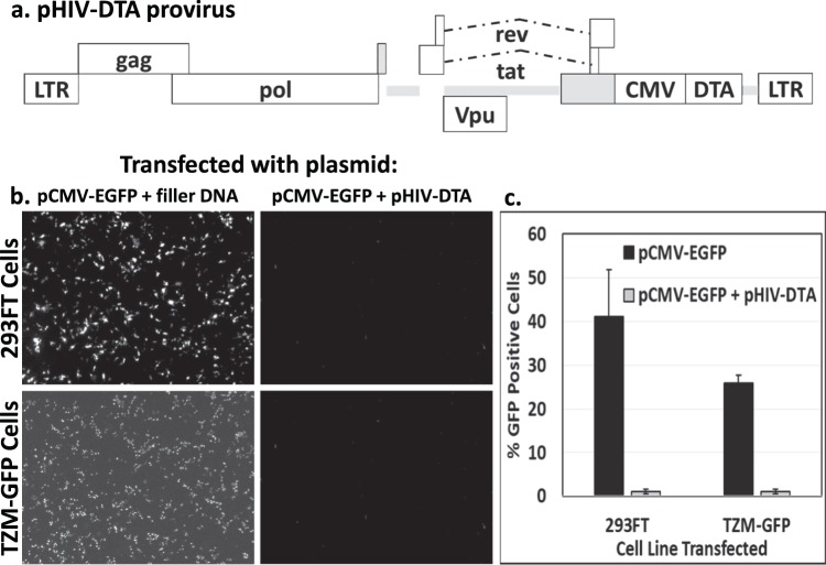
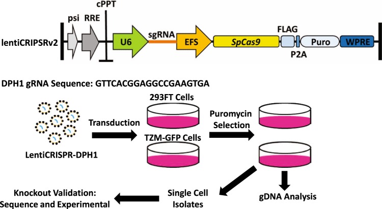
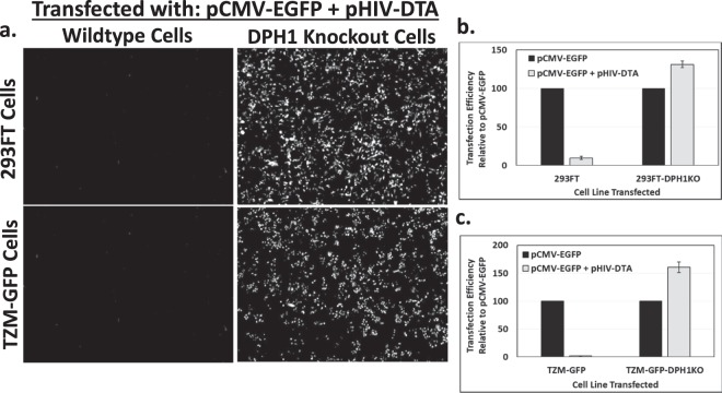
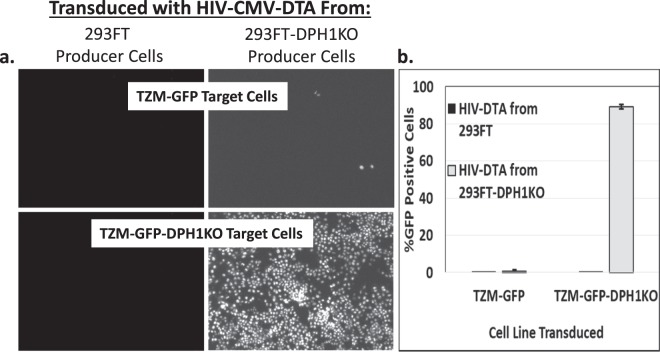
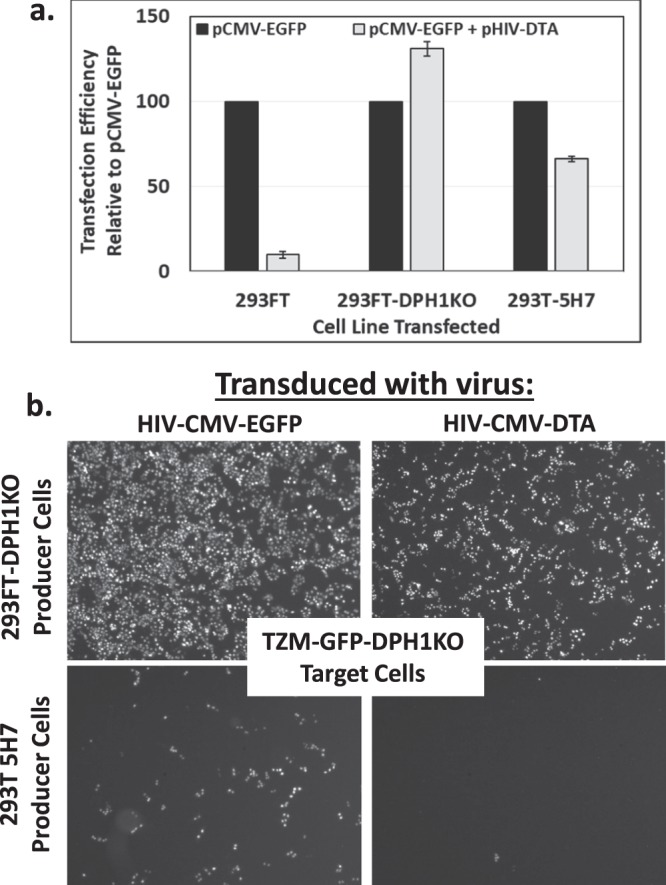
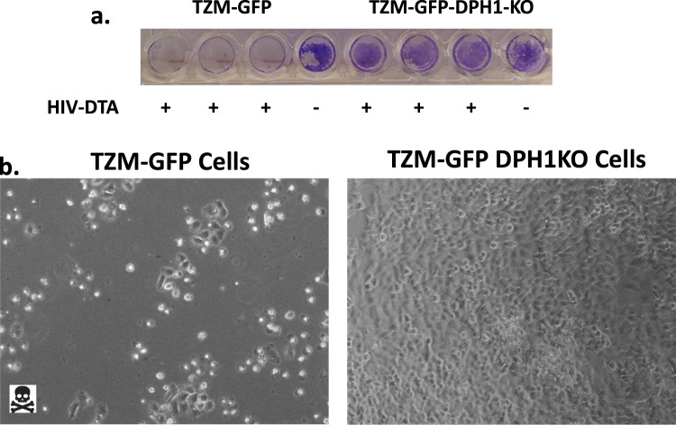
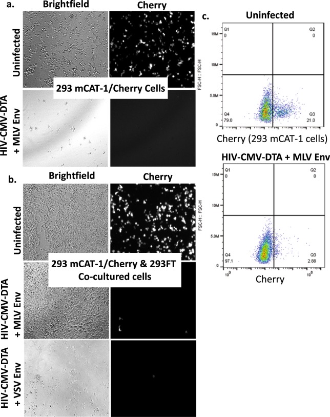
Suicide genes have been widely investigated for their utility as therapeutic agents and as tools for in vitro negative selection strategies. Several methods for delivery of suicide genes have been explored. Two important considerations for delivery are the quantity of delivered cargo and the ability to target the cargo to specific cells. Delivery using a lentiviral vector is particularly attractive due to the ability to encode the gene within the viral genome, as well as the ability to limit off-target effects by using cell type-specific glycoproteins. Here, we present the design and validation of a diphtheria toxin A (DTA)-encoding lentiviral vector expressing DTA under the control of a constituitive promoter to allow for expression of DTA in a variety of cell types, with specificity provided via selection of glycoproteins for pseudotyping of the lentiviral particles. DTA exerts its toxic activity through inhibition of eukaryotic translation elongation factor 2 (eEF2) via adenosine diphosphate (ADP)-ribosylation of a modified histidine residue, diphthamide, at His715, which blocks protein translation and leads to cell death. Thus, we also detail development of DTA-resistant cell lines, engineered through CRISPR/Cas9-mediated knockout of the diphthamide 1 (DPH1) gene, which enable both robust virus production by transfection and evaluation of DTA-expressing virus infectivity.
Conflict of interest statement
The authors declare no competing interests.
Figures
Similar articles
-
Insights into diphthamide, key diphtheria toxin effector.Toxins (Basel). 2013 May 3;5(5):958-68. doi: 10.3390/toxins5050958. Toxins (Basel). 2013. PMID: 23645155 Free PMC article.
-
Regression of prostate cancer xenografts by a lentiviral vector specifically expressing diphtheria toxin A.Cancer Gene Ther. 2003 Oct;10(10):764-70. doi: 10.1038/sj.cgt.7700629. Cancer Gene Ther. 2003. PMID: 14502229
-
Development of targeted therapy for bladder cancer mediated by a double promoter plasmid expressing diphtheria toxin under the control of H19 and IGF2-P4 regulatory sequences.J Transl Med. 2010 Dec 16;8:134. doi: 10.1186/1479-5876-8-134. J Transl Med. 2010. PMID: 21162716 Free PMC article.
-
Toward Tightly Tuned Gene Expression Following Lentiviral Vector Transduction.Viruses. 2020 Dec 11;12(12):1427. doi: 10.3390/v12121427. Viruses. 2020. PMID: 33322556 Free PMC article. Review.
-
Strategies for targeting lentiviral vectors.Curr Gene Ther. 2008 Dec;8(6):449-60. doi: 10.2174/156652308786848003. Curr Gene Ther. 2008. PMID: 19075628 Review.
Cited by
-
Virus-Free Method to Control and Enhance Extracellular Vesicle Cargo Loading and Delivery.ACS Appl Bio Mater. 2023 Mar 20;6(3):1081-1091. doi: 10.1021/acsabm.2c00955. Epub 2023 Feb 13. ACS Appl Bio Mater. 2023. PMID: 36781171 Free PMC article.
-
Hosts and Heterologous Expression Strategies of Recombinant Toxins for Therapeutic Purposes.Toxins (Basel). 2023 Dec 13;15(12):699. doi: 10.3390/toxins15120699. Toxins (Basel). 2023. PMID: 38133203 Free PMC article. Review.
-
Efficient cell death mediated by bioengineered killer extracellular vesicles.Sci Rep. 2023 Jan 19;13(1):1086. doi: 10.1038/s41598-023-28306-8. Sci Rep. 2023. PMID: 36658184 Free PMC article.
-
Toward Structurally Novel and Metabolically Stable HIV-1 Capsid-Targeting Small Molecules.Viruses. 2020 Apr 16;12(4):452. doi: 10.3390/v12040452. Viruses. 2020. PMID: 32316297 Free PMC article.
-
Design, Synthesis and Characterization of HIV-1 CA-Targeting Small Molecules: Conformational Restriction of PF74.Viruses. 2021 Mar 15;13(3):479. doi: 10.3390/v13030479. Viruses. 2021. PMID: 33804121 Free PMC article.
References
-
- Oppenheimer NJ, Bodley JW. Diphtheria toxin. Site and configuration of ADP-ribosylation of diphthamide in elongation factor 2. J Biol Chem. 1981;256:8579–8581. - PubMed
Publication types
MeSH terms
Substances
Grants and funding
LinkOut - more resources
Full Text Sources
Other Literature Sources
Miscellaneous
