Observational Study
. 2019 Jun;24(6):1-12.
doi: 10.1117/1.JBO.24.6.066008.

Detecting inflammation in rheumatoid arthritis using Fourier transform analysis of dorsal optical transmission images from a pilot study

Affiliations
Observational Study

Detecting inflammation in rheumatoid arthritis using Fourier transform analysis of dorsal optical transmission images from a pilot study

Daniel Lighter et al. J Biomed Opt. 2019 Jun.

Abstract

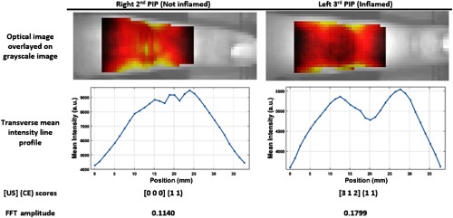
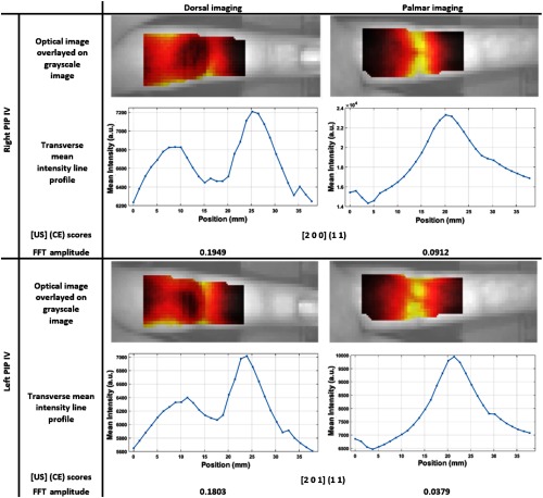
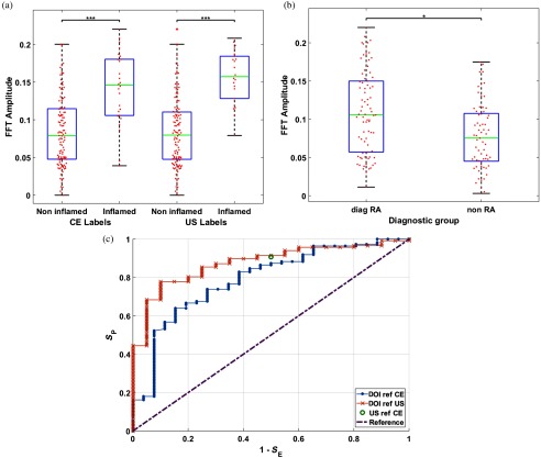
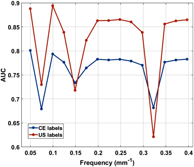
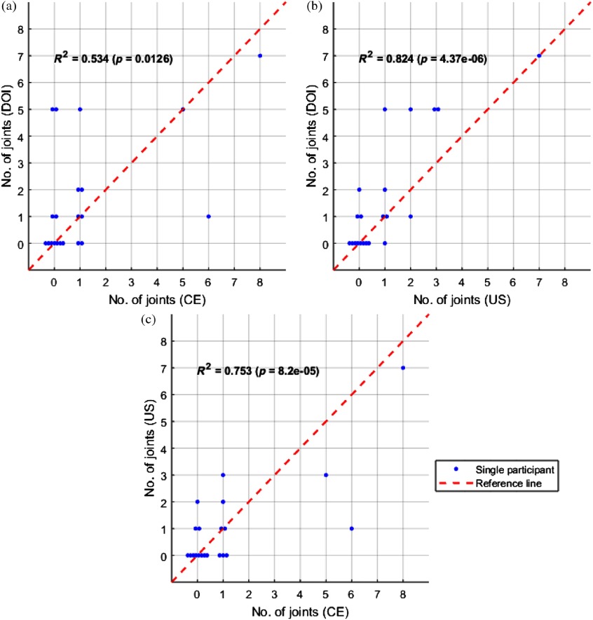
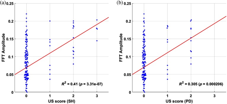
A clinical need exists for low-cost and noninvasive imaging tools capable of detecting inflammation in the joints of inflammatory arthritis patients. Previous studies have reported an optical contrast between inflamed and noninflamed joints resulting from distinct absorption and scattering properties. Accurate classification using nonocclusion-based continuous wave, transillumination imaging was limited to patient-specific changes during follow-up examination as opposed to single time-point examination, which was attributed to high intersubject variability. In distinction from previous work, optical images were acquired from the dorsal side with illumination on the palmar side and features about the spatial distribution of transmitted light along the joint were assessed using a normalized Fourier transform method. Results using this approach demonstrated an area under receiver operator curve of up to 0.888 for detecting inflammation in a pilot study involving single time-point examination of 144 joints from 21 rheumatology patients. This workflow may enable future development of clinically viable, low-cost devices for assessing inflammation in arthritis patients, without the need for cuff occlusion or comparison to baseline.

Keywords: intrinsic contrast; optical imaging; rheumatoid arthritis.


Figures

Fig. 1
Fig. 1
Illustration of the joint of an RA patient with inflammation on the dorsal side.
Fig. 2
Fig. 2
(a) 2-D schematic of system for dorsal optical transmission images acquisition. (b) Photograph of the optical imaging system setup in the rheumatology clinic. Patients placed their hand on the imaging platform and aligned their PIP joint with the source positions before acquisition.
Fig. 3
Fig. 3
Data processing steps applied to an example PIP joint, for feature extraction, with (a) grayscale image of finger with red line showing boundary of ROI defined from image mask, (b) grayscale image overlaid with transmitted DOI image from through the joint in color, (c) mean intensity across transverse of DOI image plotted for each sagittal pixel, and (d) FFT amplitude spectrum of the normalized MIT profile.
Fig. 4
Fig. 4
Simplified 2-D model of the joint and imaging setup with a single source and 27 virtual detector positions.
Fig. 5
Fig. 5
Simulation demonstrating the impact of depth of inflammation in finger from detection boundary on observed contrast in spatial profile of boundary data.
Fig. 6
Fig. 6
Two example PIP joints from the same participant. Top row: optical dorsal transmission images at 650 nm when the point source is directly under the joint, overlaid on a brightfield image of the finger. Second row: corresponding line profiles of the mean intensity in the transverse direction. Third row: reference scores [SHEFPD] (CTE CSW) for each joint. Bottom row: normalized FFT amplitude score at 0.050  mm1 frequency.
Fig. 7
Fig. 7
Two PIP joints from the same participant each with examples of both the dorsal and palmar imaged from approaches for the same joint. For each joint, (top row) optical dorsal transmission images at 650 nm when the point source is directly under the joint, overlaid on a brightfield image of the finger; (second row) corresponding line profiles of the mean intensity in the transverse direction; (third row) reference scores [SHEFPD] (CTE CSW) for each joint; and (bottom row) normalized FFT amplitude score at 0.050  mm1 frequency.
Fig. 8
Fig. 8
Example boxplots for the FFT amplitude at a frequency of 0.050  mm1, (a) comparing noninflamed against inflamed joints, labeled either using CE or US, or (b) comparing joints from the diag-RA patient group with those from the non-RA group. (c) ROC plots for DOI with either CE or US as reference labels, corresponding plot of SE and SP for US when CE is used as a reference label and the reference line corresponding to no ability to differentiate between inflamed and noninflamed joints. A marker of the sensitivity using US with CE. For each graph DOI data were collected at either 650 nm.
Fig. 9
Fig. 9
Variation of AUC in ROC analysis at all FFT frequencies for 650-nm DOI data when either US or CE were used as labels.
Fig. 10
Fig. 10
Plots comparing the number of inflamed joints on a patient level between either (a) DOI versus CE, (b) DOI versus US, or (c) US versus CE, with each marker representing a single participant. Joints were classified as inflamed for DOI using 650-nm data, with a threshold corresponding to the optimum Youden index (J=SE+SP1) established from ROC analysis.
Fig. 11
Fig. 11
Beeswarm plots with each marker representing an individual joint, comparing the relationship for US scores for (a) SH or (b) hyperaemia (power Doppler) with normalized FFT amplitude values at 0.050  mm1 frequency using 650-nm data. The red line shows the first-order polynomial fit.

References

    1. Barrett E., et al. , “The impact of rheumatoid arthritis on employment status in the early years of disease: a UK community-based study,” Rheumatology 39(12), 1403–1409 (2000).RHEUBD10.1093/rheumatology/39.12.1403 - DOI - PubMed
    1. National Audit Office, Services for People with Rheumatoid Arthritis, National Audit Office, Vol. 823, pp. 1–37 (2009).
    1. N. C. C. for Chronic Conditions et al., Rheumatoid Arthritis: National Clinical Guideline for Management and Treatment in Adults, Royal College of Physicians of London; (2009). - PubMed
    1. van der Linden M. P., et al. , “Long-term impact of delay in assessment of patients with early arthritis,” Arthritis Rheum. 62(12), 3537–3546 (2010).10.1002/art.27692 - DOI - PubMed
    1. Nell V., et al. , “Benefit of very early referral and very early therapy with disease-modifying anti-rheumatic drugs in patients with early rheumatoid arthritis,” Rheumatology 43(7), 906–914 (2004).RHEUBD10.1093/rheumatology/keh199 - DOI - PubMed

Publication types