

Giant filarial retroperitoneal cyst: a diagnostic dilemma
- PMID: 31223269
- PMCID: PMC6567645
- DOI: 10.1186/s41182-019-0164-7
Giant filarial retroperitoneal cyst: a diagnostic dilemma
Abstract
Background: Filarial infections are common in most tropical and subtropical regions of the world. Lymphatic filariasis is caused by either Wuchereria bancrofti, Brugia malayi, or Brugia timori. Extralymphatic filariasis presenting as a primary retroperitoneal mass is very rare despite filariasis being endemic in many regions of India. On review of literature, only a few isolated case reports have been described.
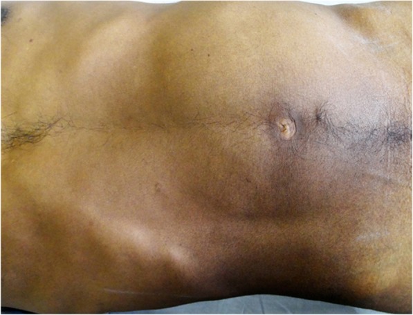
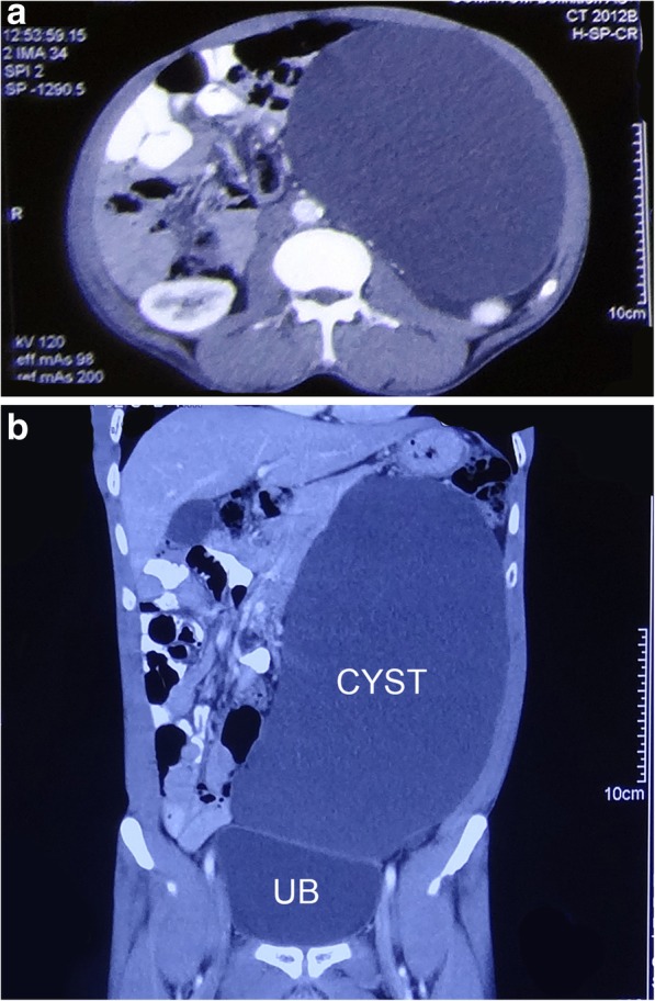
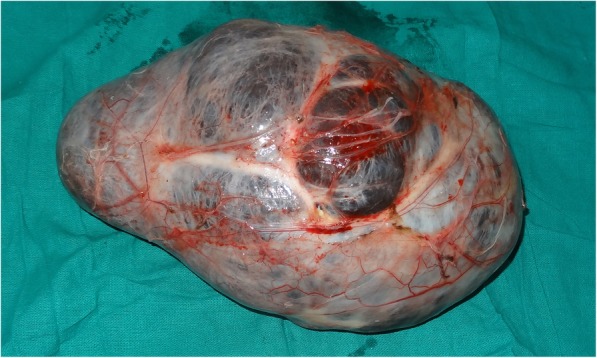
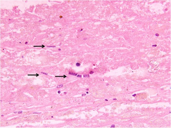
Case presentation: We report a case of a huge retroperitoneal cystic mass in a 46-year-old patient who presented with a long-standing, painless progressive abdominal swelling. On examination, there was a large, non-tender, firm swelling of size around 20 × 15 cm occupying the left upper and lower quadrant. The computed tomography of the abdomen was suggestive of thin-walled hypodense cyst of size 25.7 × 15 × 14.3 cm. Laboratory investigations and cyst aspirate were inconclusive for a definite diagnosis. On exploration, a 3-kg cystic mass was removed. The diagnosis of filarial origin was confirmed by the demonstration of microfilaria in the cyst wall and immunochromatographic test (ICT) which was positive.
Conclusion: Retroperitoneal lymphatic cyst of filarial origin is very unusual and requires a high index of suspicion if the patient is an inhabitant of an endemic area. The clinical dilemma cannot be resolved with imaging modalities alone, unless a disease-specific manifestation is there. The retroperitoneal cysts often pose a challenge in their diagnosis and management. Small cysts might respond to medical management, whereas large symptomatic cysts will require excision for the final diagnosis and treatment.
Keywords: Computed tomography; Cyst; Filariasis; Lymphatic; Retroperitoneal.
Conflict of interest statement
Competing interestsThe authors declare that they have no competing interests.
Figures
Similar articles
-
Primary retroperitoneal filariasis: a common disease of tropics with uncommon presentation and review of literature.BMJ Case Rep. 2018 Sep 26;2018:bcr2018226217. doi: 10.1136/bcr-2018-226217. BMJ Case Rep. 2018. PMID: 30257872 Free PMC article. Review.
-
Filarial huge splenomegaly dramatically regressed by anti-filarial medication: A rare clinical scenario.Intractable Rare Dis Res. 2017 Aug;6(3):215-218. doi: 10.5582/irdr.2017.01041. Intractable Rare Dis Res. 2017. PMID: 28944146 Free PMC article.
-
Role of fine needle aspiration cytology in diagnosing filarial arm cysts.BMJ Case Rep. 2013 May 17;2013:bcr2013009677. doi: 10.1136/bcr-2013-009677. BMJ Case Rep. 2013. PMID: 23687368 Free PMC article.
-
Extralymphatic Filariasis.Indian Dermatol Online J. 2023 Nov 24;15(1):92-94. doi: 10.4103/idoj.idoj_152_23. eCollection 2024 Jan-Feb. Indian Dermatol Online J. 2023. PMID: 38283023 Free PMC article.
-
Cervical Lymphatic Filariasis in a Pediatric Patient: Case Report and Database Analysis of Lymphatic Filariasis in the United States.Am J Trop Med Hyg. 2018 Jul;99(1):104-111. doi: 10.4269/ajtmh.17-0786. Epub 2018 May 24. Am J Trop Med Hyg. 2018. PMID: 29848402 Free PMC article. Review.
References
-
- Mehta RB, Gajendran V, Ananthakrishnan N, Parkash S. Retro peritoneal tumorous lesions: a clinicopathological study. Ind J Surg. 1981;43:731–742.
-
- Madhavan M, Vanaja SK, Chandra K, Reddy DJ. Atypical manifestations of filariasis in Pondycherry. Ind J Surg. 1972;34:392–394.
-
- Handfield-Jones RM. Retroperitoneal cysts: their pathology, diagnosis, and treatment. Br J Surg. 1924;12(45):119–134. doi: 10.1002/bjs.1800124515. - DOI
-
- Sharma R. Revised Kuppuswamy's Socioeconomic Status Scale: Explained and Updated. Indian Pediatr. 2017. - PubMed
-
- WHO Lymphatic Filariasis, key facts. https://www.who.int/news-room/fact-sheets/detail/lymphatic-filariasis. Accessed 7 Mar 2019.
Publication types
LinkOut - more resources
Full Text Sources