Desmoplastic infantile ganglioglioma
- PMID: 31223403
- PMCID: PMC6560944
- DOI: 10.11604/pamj.2019.32.113.12669
Desmoplastic infantile ganglioglioma
Abstract
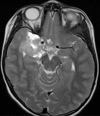
The term desmoplastic infantile ganglioglioma was coined by VandenBerg et al in 1987. In their first report these authors referred to a rare, distinct brain tumor. About 60 cases of desmoplastic infantile ganglioglioma have been described in the literature since its first description. We report a case of a 6-year-old girl who was admitted for seizure without family history. Magnetic resonance imaging scan showed a hypodense area in the right temporal region. A right temporal craniotomy was performed and the tumor was excised. The pathologic examination revealed the diagnosis of desmoplastic infantile ganglioglioma.
Keywords: Tumor; central nervous system; infantile desmoplastic ganglioglioma.
Conflict of interest statement
The authors declare no competing interest.
Figures


References
-
- VandenBerg SR, May EE, Rubinstein LJ, Herman MM, Perentes E, Vinores S, et al. Desmoplastic supratentorial neuroepithelial tumors of infancy with divergent differentiation potential (“desmoplastic infantile gangliogliomas”) Report on 11 cases of a distinctive embryonal tumor with favorable prognosis. Journal of neurosurgery. 1987;66(1):58–71. - PubMed
-
- Iwami K-i. Desmoplastic infantile ganglioglioma. Child's Nervous System. 2007;23(6):619–20. - PubMed
-
- Craver RD, Nadell J, Nelson JS. Desmoplastic infantile ganglioglioma. Pediatr Dev Pathol. 1999 Nov-Dec;2(6):582–7. - PubMed
-
- Prayson RA, Cohen ML, Kleinschmidt-De Masters B. Brain Tumors. 2009.
Publication types
MeSH terms
LinkOut - more resources
Full Text Sources
Medical
