. 2019 Jun 19;5(6):eaav9946.
doi: 10.1126/sciadv.aav9946. eCollection 2019 Jun.

Sortilin gates neurotensin and BDNF signaling to control peripheral neuropathic pain

Affiliations

Sortilin gates neurotensin and BDNF signaling to control peripheral neuropathic pain

Mette Richner et al. Sci Adv. .

Abstract

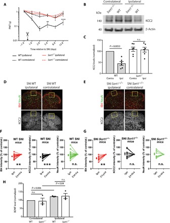
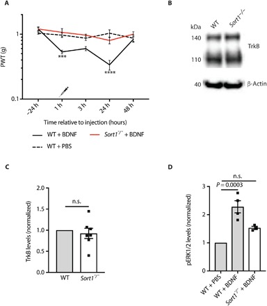
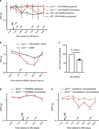
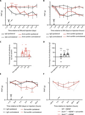
Neuropathic pain is a major incurable clinical problem resulting from peripheral nerve trauma or disease. A central mechanism is the reduced expression of the potassium chloride cotransporter 2 (KCC2) in dorsal horn neurons induced by brain-derived neurotrophic factor (BDNF), causing neuronal disinhibition within spinal nociceptive pathways. Here, we demonstrate how neurotensin receptor 2 (NTSR2) signaling impairs BDNF-induced spinal KCC2 down-regulation, showing how these two pathways converge to control the abnormal sensory response following peripheral nerve injury. We establish how sortilin regulates this convergence by scavenging neurotensin from binding to NTSR2, thus modulating its inhibitory effect on BDNF-mediated mechanical allodynia. Using sortilin-deficient mice or receptor inhibition by antibodies or a small-molecule antagonist, we lastly demonstrate that we are able to fully block BDNF-induced pain and alleviate injury-induced neuropathic pain, validating sortilin as a clinically relevant target.


Figures

Fig. 1
Fig. 1. KCC2 down-regulation is prevented in sortilin-deficient mice.
(A) Paw withdrawal threshold (PWT) to tactile stimuli of ipsilateral versus contralateral sides of WT and Sort1−/− mice before and after SNI (day 0). *P < 0.02, **P < 0.009, and ****P < 0.0001; n.s., not significant; n = 7 to 8, two-way repeated measures (RM) analysis of variance (ANOVA) with post hoc Tukey’s test [F(3,26) = 31.52, P < 0.0001], means ± SEM. (B) Representative Western blot of KCC2 in L3-L5 SDH 6 days after SNI. (C) KCC2 levels in L3-L5 SDH quantified by Western blot and normalized to WT contralateral 6 days after SNI. n = 6, one-way RM ANOVA with post hoc Tukey’s test [F(1.997,9.985) = 15.06, P = 0.001], means ± SEM. (D and E) IHC analysis showing IB4, NeuN, and KCC2 expression in the ipsilateral and contralateral SDH of WT and Sort1−/− mice. Scale bar, 100 μm. (F and G) Comparisons of average pixel intensity are shown across SNI animals of WT versus Sort1−/− mice in the region of interest (ROI). Nerve injury resulted in decreased IB4 intensity in the ROI in WT mice (contralateral versus ipsilateral: paired t test, t = 3.749; df = 18, P = 0.0015; n = 19) as in Sort1−/− mice (contralateral versus ipsilateral: paired t test, t = 4; df = 8, P = 0.004; n = 9). Nerve injury caused the down-regulation of KCC2 expression in the dorsal horn of WT mice but not in Sort1−/− mice [contralateral versus ipsilateral: (WT mice) paired t test, t = 6.24; df = 18, P < 0.0001; n = 19; and (Sort1−/− mice) t = 0.2093; df = 8, P = 0.839; n = 9]. No loss of neurons, measured as the difference in the average NeuN immunostaining intensities, was observed between ipsilateral and contralateral sides in both WT and Sort1−/− mice [contralateral versus ipsilateral: (WT mice) paired t test, t = 1.206; df = 18, P = 0.2436; n = 19; and (Sort1−/− mice) t = 0.3838; df = 8, P = 0.7111; n = 9]. **P < 0.01 and ***P < 0.0001; intensity units (i.u.) are shown as means ± SEM. (H) BDNF levels 6 days after SNI in L3-L5 SDH relative to WT contralateral [n = 3, pooled samples from eight mice for each run, paired t test within genotype (WT: t = 13.42, df = 2; Sort1−/−: t = 4.62, df = 2) and unpaired t test between genotypes (means ± SEM)].
Fig. 2
Fig. 2. Spinal BDNF signaling is altered in the absence of sortilin.
(A) PWT to tactile stimuli of WT and Sort1−/− mice before and after intrathecal BDNF or vehicle injection. ***P < 0.0009 and ****P < 0.0001, versus WT + vehicle; n = 6, two-way RM ANOVA with post hoc Dunnett’s test [F(2,15) = 10.96, P < 0.0012], means ± SEM. (B) Representative Western blot of TrkB in L3-L5 SDH. (C) TrkB levels in L3-L5 SDH in naïve WT and Sort1−/− mice quantified by Western blotting. n = 7, t test, means ± SEM. (D) pERK1/2 levels in L3-L5 SDH 3 hours after intrathecal phosphate-buffered saline (PBS) or BDNF injection at spinal L3-L5. n = 3 to 4, one-way ANOVA with post hoc Dunnett’s test [F(2,8) = 22.58, P = 0.0003), means ± SEM.
Fig. 3
Fig. 3. Sortilin deletion unmasks NTSR2 signaling.
(A) PWT to tactile stimuli of WT and Sort1−/− mice before and after SNI and intrathecal SR142948A injection. ***P < 0.0004 and ****P < 0.0001, versus the contralateral side of Sort1−/− mice; n = 6 to 8, two-way RM ANOVA with post hoc Dunnett’s test [F(3,24) = 35.53, P < 0.0001], means ± SEM, with syringes representing double injections per day. (B) PWT to tactile stimuli of Sort1−/− mice before and after intrathecal BDNF ± SR142948A injection. **P < 0.009 and ***P < 0.0008; n = 6, two-way RM ANOVA with post hoc Sidak’s test [F(1,10) = 10.49, P = 0.0089], means ± SEM, with the syringe representing double intraperitoneal SR142947A injections on days −2, −1, and 0 and one intrathecal BDNF injection on day 0, 1 hour before testing. (C) KCC2 levels in ipsilateral L3-L5 SDH of Sort1−/− mice with and without intrathecal SR142948A injection 6 days after SNI. n = 3, paired t test, means ± SEM. (D) PWT to tactile stimuli of Sort1−/− mice before and after SNI and intraperitoneal SR48692. n = 6, two-way RM ANOVA with post hoc Dunnett’s test, means ± SEM, with syringes representing double injections per day. (E) PWT to tactile stimuli of Sort1−/− mice before and after SNI and intraperitoneal levocabastine. ***P < 0.0003 and ****P < 0.0001; n = 7, two-way RM ANOVA with post hoc Dunnett’s test [F(3,24) = 39.06, P < 0.0001], means ± SEM, with syringes representing single injections.
Fig. 4
Fig. 4. The extracellular domain of sortilin is sufficient for BDNF/TrkB signaling.
(A) PWT to tactile stimuli of WT mice before and after SNI and intrathecal anti-sortilin antibodies or immunoglobulin G (IgG) injection at day 0. ****P < 0.0001 for ipsilateral anti-sortilin versus ipsilateral IgG; n = 6, two-way RM ANOVA with post hoc Dunnett’s test [F(3,20) = 48.09, P < 0.0001], means ± SEM. (B) PWT to tactile stimuli of WT mice before and after SNI (day 0) and intrathecal anti-sortilin antibodies or IgG injection 6 days after SNI, means ± SEM. (C) Percentage of anti-allodynia by anti-sortilin 1, 2, and 3 hours versus pre-injection baseline (average of days 2 and 6 of the same mouse). *P < 0.03 and **P < 0.001; n = 5, one-way RM ANOVA with post hoc Dunnett’s test [F(3,12) = 9.691, P = 0.0016], means ± SEM. (D) Percentage of anti-allodynia by IgG 1, 2, and 3 hours versus average of days 2 and 6 of the same mouse. n = 5, one-way RM ANOVA with post hoc Dunnett’s test, means ± SEM. (E) PWT to tactile stimuli of Sort1−/− mice before and after SNI (day 0) and intrathecal sol-sortilin or IgG injection (0 hours). *P < 0.02 and ****P < 0.0001, versus the ipsilateral side of intrathecal IgG; n = 4 to 7, two-way RM ANOVA with post hoc Dunnett’s test [F(3,18) = 15.17, P < 0.0001], means ± SEM. (F) PWT to tactile stimuli of WT and Sort1−/− mice before and after intrathecal BDNF ± sol-sortilin injection. ****P < 0.0001 versus Sort1−/− + BDNF injection; n = 6 to 12, two-way RM ANOVA with post hoc Dunnett’s test [F(2,23) = 10.85, P = 0,0005], means ± SEM.
Fig. 5
Fig. 5. Pharmacological inhibition of sortilin prevents mechanical allodynia.
(A) PWT to tactile stimuli of WT and Sort1−/− mice before and after SNI and intrathecal AF38469 (active) or AF38540 (inactive) injection. ****P < 0.0001 versus the ipsilateral side of WT + AF38540; n = 13 to 17, two-way RM ANOVA with post hoc Dunnett’s test [F(5,66) = 17.75, P < 0.0001], means ± SEM. (B) PWT to tactile stimuli of WT mice after intrathecal AF38469 (active) or AF38540 (inactive) injection 2 days after SNI. (C) Percentage of anti-allodynia by AF38469 1, 2, and 3 hours versus pre-injection baseline (day 2 of the same mouse). ***P < 0.0007; n = 9, one-way RM ANOVA with post hoc Dunnett’s test [F(3,24) = 8.597, P = 0.0005], means ± SEM. (D) Percentage of anti-allodynia by AF38540 1, 2, and 3 hours versus pre-injection baseline (day 2 of the same mouse). n = 6, one-way RM ANOVA with post hoc Dunnett’s test, means ± SEM. (E) PWT to tactile stimuli of WT mice before and after intrathecal BDNF ± AF38469 (active) injection. *P < 0.03, ***P < 0.0004, and ****P < 0.0001; n = 6 to 7, two-way RM ANOVA with post hoc Sidak’s test [F(1,11) = 19, P = 0.0011], means ± SEM. (F) PWT to tactile stimuli of hum-sortilin-KI mice after intrathecal AF38469 (active) or AF38540 (inactive) injection 2 days after SNI. ****P < 0.0001 versus ipsilateral side with AF38540; n = 8, two-way RM ANOVA with post hoc Dunnett’s test [F(3,28) = 45.67, P < 0.0001, means ± SEM.

Similar articles

Cited by

References

    1. van Hecke O., Austin S. K., Khan R. A., Smith B. H., Torrance N., Neuropathic pain in the general population: A systematic review of epidemiological studies. Pain 155, 654–662 (2014). - PubMed
    1. Coull J. A. M., Boudreau D., Bachand K., Prescott S. A., Nault F., Sík A., De Koninck P., De Koninck Y., Trans-synaptic shift in anion gradient in spinal lamina I neurons as a mechanism of neuropathic pain. Nature 424, 938–942 (2003). - PubMed
    1. Lu Y., Zheng J., Xiong L., Zimmermann M., Yang J., Spinal cord injury-induced attenuation of GABAergic inhibition in spinal dorsal horn circuits is associated with down-regulation of the chloride transporter KCC2 in rat. J. Physiol. 586, 5701–5715 (2008). - PMC - PubMed
    1. Doyon N., Vinay L., Prescott S. A., De Koninck Y., Chloride regulation: A dynamic equilibrium crucial for synaptic inhibition. Neuron 89, 1157–1172 (2016). - PubMed
    1. Coull J. A. M., Beggs S., Boudreau D., Boivin D., Tsuda M., Inoue K., Gravel C., Salter M. W., De Koninck Y., BDNF from microglia causes the shift in neuronal anion gradient underlying neuropathic pain. Nature 438, 1017–1021 (2005). - PubMed

Publication types

LinkOut - more resources