. 2019 Jun 19;5(6):eaax2650.
doi: 10.1126/sciadv.aax2650. eCollection 2019 Jun.

Structure of the saxiphilin:saxitoxin (STX) complex reveals a convergent molecular recognition strategy for paralytic toxins

Affiliations

Structure of the saxiphilin:saxitoxin (STX) complex reveals a convergent molecular recognition strategy for paralytic toxins

Tien-Jui Yen et al. Sci Adv. .

Abstract

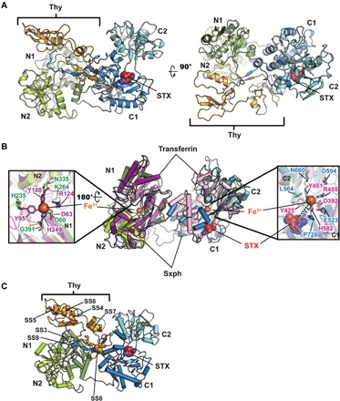
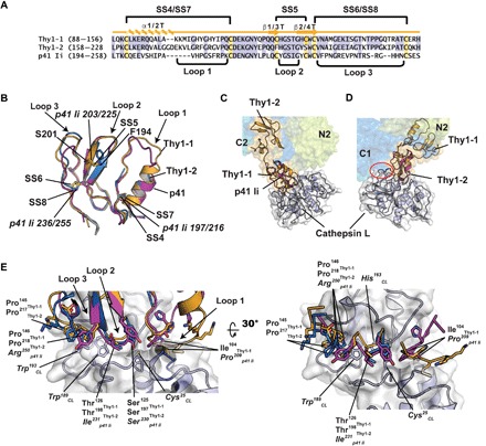
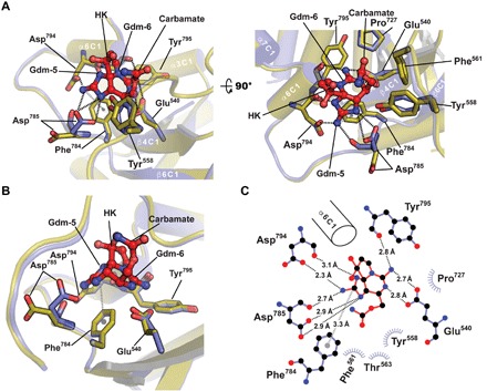
Dinoflagelates and cyanobacteria produce saxitoxin (STX), a lethal bis-guanidinium neurotoxin causing paralytic shellfish poisoning. A number of metazoans have soluble STX-binding proteins that may prevent STX intoxication. However, their STX molecular recognition mechanisms remain unknown. Here, we present structures of saxiphilin (Sxph), a bullfrog high-affinity STX-binding protein, alone and bound to STX. The structures reveal a novel high-affinity STX-binding site built from a "proto-pocket" on a transferrin scaffold that also bears thyroglobulin domain protease inhibitor repeats. Comparison of Sxph and voltage-gated sodium channel STX-binding sites reveals a convergent toxin recognition strategy comprising a largely rigid binding site where acidic side chains and a cation-π interaction engage STX. These studies reveal molecular rules for STX recognition, outline how a toxin-binding site can be built on a naïve scaffold, and open a path to developing protein sensors for environmental STX monitoring and new biologics for STX intoxication mitigation.


Figures

Fig. 1
Fig. 1. Sxph structure.
(A) R. catesbeiana Sxph:STX: complex ribbon diagram. Domains are indicated and are colored as follows: N1 (smudge), N2 (limon), thyroglobulin (Thy; bright orange), C1 (marine), and C2 (cyan). STX (red) is shown as space filling. (B) Superposition of Sxph and rabbit transferrin [Protein Data Bank (PDB): 1JNF] (32). Transferrin N-lobe and C-lobe are colored purple and pink, respectively. Sxph Thy1 repeats are not shown. Insets show transferrin Fe3+ ligands and Sxph equivalents as sticks. N domain: transferrin (purple) and Sxph (green); C domain: transferrin (pink) and Sxph (blue). STX (red) is shown as space filling. Right hand inset shows distance between the STX center and transferrin Fe3+. (C) Cartoon diagram showing unique Sxph disulfide bonds in space filling representation: SS3 (Cys27 to Cys417) SS4 (Cys91 to Cys111), SS5 (Cys122 to Cys129), SS6 (Cys131 to Cys153), SS7 (Cys161 to Cys183), SS8 (Cys203 to Cys225), and SS9 (Cys234 to Cys825). Colors and labels are the same as in (A).
Fig. 2
Fig. 2. Comparison of Sxph Thy1-1 and Thy1-2 with p41 Ii.
(A) Sequence comparison. Thy1-1 and Thy1-2 secondary structure elements and disulfide bonds are indicated. Cysteines and conserved residues are highlighted yellow and blue, respectively. (B) Cartoon diagram superposition of Thy1-1 (light orange), Thy1-2 (marine), and p41 Ii (magenta) (PDB: 1ICF) (38). Disulfide bonds (italics) and select residues are labeled. Thy1-1 and Thy1-2 have root mean square deviation of Cα position (RMSD) = 0.61 and 0.64 Å over 43 and 42 residues, respectively. p41 Ii has RMSD = 0.57 Å over 53 residues of Sxph Thy1-1 and Thy1-2. (C and D) Superposition of Sxph on the p41 Ii:cathespin L complex (PDB: 1ICF) (38) using the (C) Thy1-1 and (D) Thy1-2 domains. In (D), red oval indicates cathepsin L and Sxph C1 clash. Sxph colors are the same as in Fig. 1A. (E) Superposition of Sxph Thy1-1 (light orange), Thy1-2 (marine), and p41 Ii (magenta) in the context of the p41 Ii:cathepsin L interface.
Fig. 3
Fig. 3. Sxph STX-binding site.
(A) Apo-Sxph (olive) and STX-bound Sxph (slate) superposition cartoon diagram. STX-interacting residues are shown as sticks. Key secondary structure elements are labeled. Black and gray dashed lines indicate hydrogen bond networks and the cation-π interaction, respectively. STX is shown as red sticks. Gdm-5, Gdm-6, and HK indicate the five- and six-membered guanidinium rings and hemiketal, respectively. (B) STX-binding site highlighting the cation-π interaction (gray) and Asp785 movement. (C) LIGPLOT diagram of the STX-binding site. α6C1 is shown for orientation.
Fig. 4
Fig. 4. Sxph STX-binding site and transferrin proto-pocket.
(A and B) Superposition of the Sxph (marine) STX-binding site with (A) Fe3+-bound rabbit serum transferrin (PDB: 1JNF) (41) (pink) and (B) apo-human serum transferrin (PDB: 2HAU) (32) (gray). STX (red) is shown as sticks. Select residues are shown. Blue labels indicate Sxph residues. Orange arrows indicate changes between transferrin and Sxph. (C to E) Transferrin proto-pocket and Sxph STX-binding pocket comparisons. (C) to (E) show apo-transferrin (pink), transferrin (gray), and Sxph (marine) surfaces, respectively. In (C) and (D), labels indicate Sxph residues that break through the transferrin surface. Red circle highlights the STX-binding site. STX is shown as space filling. Sxph surface is colored by atom type, where red and blue denote oxygen and nitrogen, respectively.
Fig. 5
Fig. 5. Sxph and NaVs share STX recognition strategies.
(A) NaVPaS:STX (PDB: 6a91) (29) and Sxph:STX STX-binding site superposition. NaVPaS is shown as a cartoon viewed from the central channel cavity. Pore domains are colored as follows: DI, green; DII, orange; DIII, yellow; and DIV, pink. STX coordinating and selectivity filter “DEKA” motif (white) residues are shown as sticks. Sxph STX-binding site side chains are blue. STX from Sxph:STX (red) and NaVPaS:STX (cyan) are superposed. (B) Closeup view of the STX-binding sites from (A). (C) Diagram of the NaVPaS:STX interactions. (D) Diagram of the Sxph:STX interactions. (E) Comparison of common STX interactions for Sxph (blue), NaVPaS (green), and NaV1.7 (magenta). STX from the Sxph:STX complex (red), NaVPaS:STX complex (cyan), and NaV1.7:STX complex (violet) are indicated. (C) and (D) were generated using LIGPLOT (67) and a 3.35-Å cutoff. Hydrogen bonding networks (black dashed lines) and cation-π interactions (gray dashed lines) are indicated. (D) is the same as Fig. 3C.

References

    1. Thottumkara A. P., Parsons W. H., Du Bois J., Saxitoxin. Angew. Chem. Int. Ed. Engl. 53, 5760–5784 (2014). - PubMed
    1. Duran-Riveroll L. M., Cembella A. D., Guanidinium toxins and their interactions with voltage-gated sodium ion channels. Mar. Drugs 15, 303 (2017). - PMC - PubMed
    1. B. Hille, Ion Channels of Excitable Membranes (Sinauer Associates Inc., ed. 3, 2001).
    1. Su Z., Sheets M., Ishida H., Li F., Barry W. H., Saxitoxin blocks L-type ICa. J. Pharmacol. Exp. Ther. 308, 324–329 (2004). - PubMed
    1. Wang J., Salata J. J., Bennett P. B., Saxitoxin is a gating modifier of HERG K+ channels. J. Gen. Physiol. 121, 583–598 (2003). - PMC - PubMed

Publication types