

A mathematical model for the first derivative wave analysis of the volumetric capnogram from the perspective of erythrocyte motion profiles
- PMID: 31223667
- PMCID: PMC6562574
- DOI: 10.1016/j.heliyon.2019.e01824
A mathematical model for the first derivative wave analysis of the volumetric capnogram from the perspective of erythrocyte motion profiles
Abstract
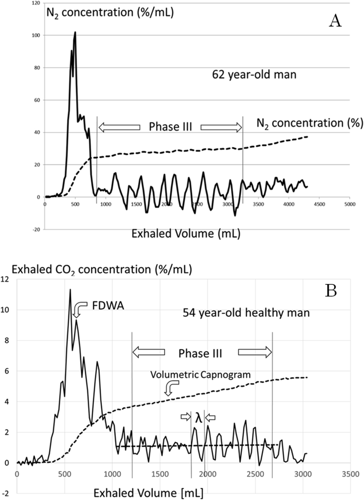
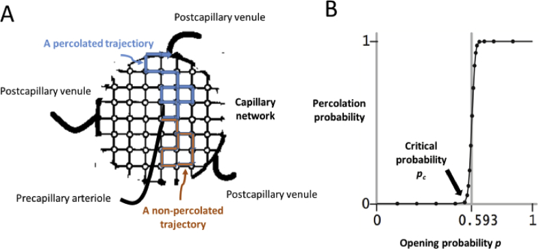
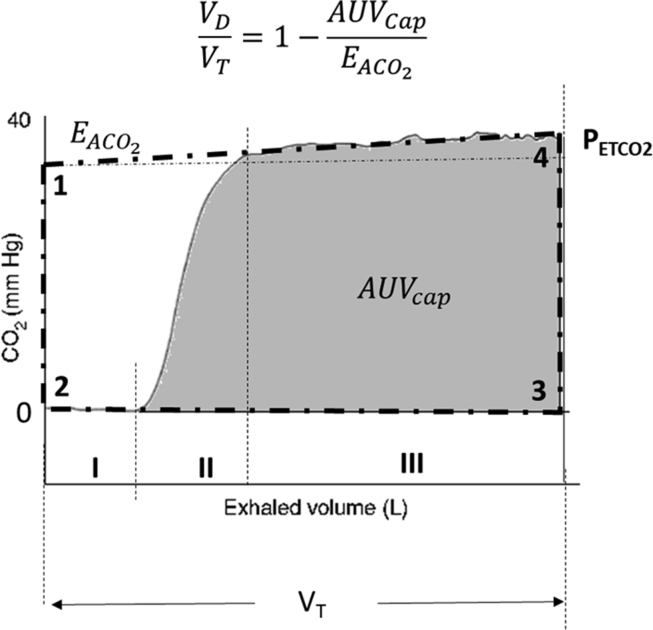
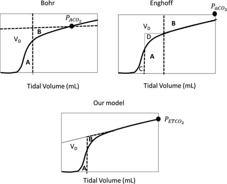
Current trends in monitoring system are leading to the adoption of volumetric capnogram (Vcap). The first derivative wave analysis (FDWA) of Vcap represented the cardiogenic oscillations (CarO) as a propagated wave and the slope of phase III (SIII) as a constant. Until today the genesis of CarO and SIII is however under debate. In this study, we defined motion profiles of erythrocytes in the pulmonary parenchyma as pulsated-run and random-walk, on the basis of which we obtained a new mathematical expression describing FDWA of Vcap. The mathematical model of Vcap provided theoretical explanation concerned with motion profiles of erythrocytes about the genesis of CarO and SIII. As the results, the mathematical model predicted the close relationship between SIII and the transfer factor of carbon monoxide, which will be used for estimating validity of this mathematical model. In addition, the velocity of propagated wave in the phase III was suggested as a new physiological variable to estimate elastic properties of pulmonary arterioles, and a new measuring method of VD was proposed based on the theoretical reason, as well. Clinical investigations of the new VD to test its efficacy of monitoring are needed.
Keywords: Cardiogenic oscillation; Dead space; First derivative wave analysis; Mathematical biosciences; Phase III slope; Physiology; Pulsated-run; Random-walk; Volumetric capnogram.
Figures
Similar articles
-
A simplified 4-parameter model of volumetric capnograms improves calculations of airway dead space and slope of Phase III.J Clin Monit Comput. 2020 Dec;34(6):1265-1274. doi: 10.1007/s10877-019-00451-4. Epub 2019 Dec 23. J Clin Monit Comput. 2020. PMID: 31872310
-
Capnogram slope and ventilation dead space parameters: comparison of mainstream and sidestream techniques.Br J Anaesth. 2016 Jul;117(1):109-17. doi: 10.1093/bja/aew127. Br J Anaesth. 2016. PMID: 27317710 Free PMC article.
-
Volumetric capnography: the time has come.Curr Opin Crit Care. 2014 Jun;20(3):333-9. doi: 10.1097/MCC.0000000000000095. Curr Opin Crit Care. 2014. PMID: 24785676 Review.
-
Assessment of dead-space ventilation in patients with acute respiratory distress syndrome: a prospective observational study.Crit Care. 2016 May 5;20(1):121. doi: 10.1186/s13054-016-1311-8. Crit Care. 2016. PMID: 27145818 Free PMC article.
-
Using the features of the time and volumetric capnogram for classification and prediction.J Clin Monit Comput. 2017 Feb;31(1):19-41. doi: 10.1007/s10877-016-9830-z. Epub 2016 Jan 18. J Clin Monit Comput. 2017. PMID: 26780902 Review.
Cited by
-
Accelerating the Diagnosis of Pandemic Infection Based on Rapid Sampling Algorithm for Fast-Response Breath Gas Analyzers.Sensors (Basel). 2024 Sep 24;24(19):6164. doi: 10.3390/s24196164. Sensors (Basel). 2024. PMID: 39409204 Free PMC article.
References
-
- Broadbent S.R., Hammersley J.M. Percolation processes. Math. Proc. Camb. Philos. Soc. 2008;53(03) 629-541.
-
- Davis P.J., Polansky I. Differences. In §25 (Numerical interporation, differentiation and integration) In: Abramowitz M., Stegun I.A., editors. Handbook of Mathematical Functions with Formulas, Graphs, and Mathematical Tables, 9th Printing. Dover; New York: 1972. pp. 877–878.
-
- Fowler W.S. Lung function studies. II. The respiratory dead space. Am. J. Physiol. 1948;154:405–416. - PubMed
LinkOut - more resources
Full Text Sources
Research Materials
