Case Reports
. 2019 Mar 2:3:100026.
doi: 10.1016/j.wnsx.2019.100026. eCollection 2019 Jul.

Distal Parent Vessel Occlusion of 2 Superior Cerebellar Artery Fusiform Aneurysms: Report of 2 Cases and Literature Review

Affiliations
Case Reports

Distal Parent Vessel Occlusion of 2 Superior Cerebellar Artery Fusiform Aneurysms: Report of 2 Cases and Literature Review

Luis C Ascanio et al. World Neurosurg X. .

Abstract

Background: Fusiform superior cerebellar artery (SCA) aneurysms are rare, and their management represents a technical challenge. In complex aneurysms, endovascular parent vessel occlusion of the SCA may be a treatment option. Here, we present 2 cases of fusiform SCA aneurysms, 1 ruptured and 1 unruptured, as well as our institution's management with parent vessel occlusion. We also provide a review of the literature.

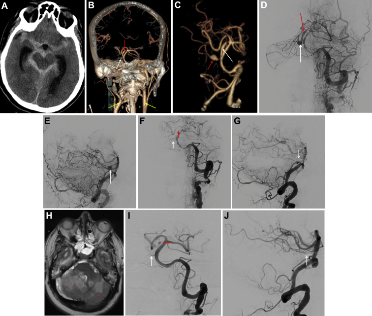
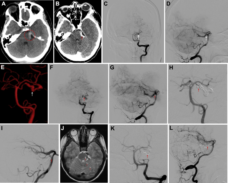
Cases description: Case 1: A 42-year-old male was transferred from an outside hospital with subarachnoid hemorrhage. On admission, the patient had a Glasgow Coma Scale score of 8, a Hunt and Hess grade 4, and a Fisher grade 4. A diagnostic angiogram demonstrated a right SCA fusiform lesion with proximal and distal dilatations of 1.45 mm and 5.35 mm long, respectively, likely representing a single dissecting pseudoaneurysm. The distal dilatation was coiled, resulting in parent vessel occlusion. The patient recovered clinically and was discharged in stable condition.Case 2: A 27-year-old female was transferred from an outside hospital due to a brainstem stroke. A diagnostic angiogram revealed an S2/S3 segment left SCA fusiform lesion, likely representing a dissecting aneurysm. The patient was neurologically intact at admission and managed conservatively. At the 2-month follow-up angiogram, the dissection had extended along the length of the SCA. Consequently, the patient underwent coil embolization of the distal left SCA. At the 6-month follow-up, the vessel remained obliterated and the patient's neurologic status had improved.

Conclusions: Endovascular coil embolization of fusiform SCA aneurysms offers a reasonable and safe treatment approach.

Keywords: Aneurysm; CT, Computed tomography; Cerebrovascular; Fusiform; OSH, Outside hospital; S2, Lateral pontomesencephalic segment; SAH, Subarachnoid hemorrhage; SCA, Superior cerebellar artery; Subarachnoid hemorrhage; Superior cerebellar artery.


Figures

Figure 1
Figure 1
Admission computed tomography (CT) shows subarachnoid hemorrhage around the basilar cisterns (A). Coronal (B) projection of head and neck CT angiography showing bilateral extracranial carotid dissection (yellow arrows) and the distal (white arrow) and proximal (red arrow) portion of a fusiform right superior cerebellar artery aneurysm. A 3-dimensional reconstruction of basilar artery CT angiography (C) shows the distal (white arrow) and proximal (red arrow) portion of the pseudoaneurysm. Anteroposterior (AP) (D) and lateral (E) projections from the preprocedure angiogram shows the distal (white arrow) and proximal (red arrow) portions of the aneurysm. AP (F) and lateral (G) projections from the postprocedure angiogram shows coil embolization and occlusion of the distal portion of the aneurysm (white arrow) and blood flow reduction to the proximal portion of the aneurysm (red arrow). Bilateral superior cerebellar infarcts (red arrows) observed on brain magnetic resonance imaging 8 days after treatment (H). A 2-months’ follow-up, angiogram revealed complete obliteration of the distal portion of the aneurysm on AP (I) and lateral (J) projections (white arrow), as well as the parent vessel with persistent flow to the proximal portion of the aneurysm (red arrow).
Figure 2
Figure 2
(A) Head computed tomography (CT) from an outside hospital revealing a hyperintensity in the left parapontine region of the brainstem (red circle). (B) Head CT angiography shows ectasia of the perimesencephalic portion of the left superior cerebellar artery (SCA) (red arrow). Admission angiogram revealed a dissection along the S2 and cerebellomesoencephalic segment segments (white arrow) of the left SCA in anteroposterior (AP) (C), lateral (D), and virtual reconstruction (E). A 2 months' follow-up, an angiogram showed extension of the previously identified dissection to the S1 and cortical segment segments on AP (F,red arrow) and lateral projections (G,red arrow). The patient underwent elective endovascular treatment by coiling the distal left SCA and leaving patent the proximal left SCA (H and I,red arrow). A 5 days' posttreatment brain T2 magnetic resonance image shows several punctate areas of diffusion abnormality in the left midbrain and left superior cerebellum suggestive of acute infarction (J,red arrows). A 6-months’ follow-up angiogram revealed complete obliteration of the left SCA (K and L,red arrow).

Similar articles

Cited by

References

    1. Awad A.J., Mascitelli J.R., Haroun R.R., De Leacy R.A., Fifi J.T., Mocco J. Endovascular management of fusiform aneurysms in the posterior circulation: the era of flow diversion. Neurosurg Focus. 2017;42:E14. - PubMed
    1. Park S.H., Yim M.B., Lee C.Y., Kim E., Son E.I. Intracranial fusiform aneurysms: it's pathogenesis, clinical characteristics and managements. J Kor Neurosurg Soc. 2008;44:116–123. - PMC - PubMed
    1. Lubicz B., Leclerc X., Gauvrit J.Y., Lejeune J.P., Pruvo J.P. Endovascular treatment of peripheral cerebellar artery aneurysms. AJNR Am J Neuroradiol. 2003;24:1208–1213. - PMC - PubMed
    1. Chaloupka J.C., Putman C.M., Awad I.A. Endovascular therapeutic approach to peripheral aneurysms of the superior cerebellar artery. AJNR Am J Neuroradiol. 1996;17:1338–1342. - PMC - PubMed
    1. Kang I.H., Malla H.P., Lee S.H., Park C.K., Choi S.K. Revascularization as treatment of a ruptured fusiform aneurysm at the cortical segment of the superior cerebellar artery: a case report and literature review. J Neurol Surg A Cent Eur Neurosurg. 2017;78:302–305. - PubMed

Publication types

LinkOut - more resources