. 2019 Oct;235(4):716-724.
doi: 10.1111/joa.13032. Epub 2019 Jun 21.

Somite development in the avian tail

Affiliations

Somite development in the avian tail

Margarethe Draga et al. J Anat. 2019 Oct.

Abstract

Somites are epithelial segments of the paraxial mesoderm. Shortly after their formation, the epithelial somites undergo extensive cellular rearrangements and form specific somite compartments, including the sclerotome and the myotome, which give rise to the axial skeleton and to striated musculature, respectively. The dynamics of somite development varies along the body axis, but most research has focused on somite development at thoracolumbar levels. The development of tail somites has not yet been thoroughly characterized, even though vertebrate tail development has been intensely studied recently with respect to the termination of segmentation and the limitation of body length in evolution. Here, we provide a detailed description of the somites in the avian tail from the beginning of tail formation at HH-stage 20 to the onset of degeneration of tail segments at HH-stage 27. We characterize the formation of somite compartment formation in the tail region with respect to morphology and the expression patterns of the sclerotomal marker gene paired-box gene 1 (Pax1) and the myotomal marker genes MyoD and myogenic factor 5 (Myf5). Our study gives insight into the development of the very last segments formed in the avian embryo, and provides a basis for further research on the development of tail somite derivatives such as tail vertebrae, pygostyle and tail musculature.

Keywords: chicken; embryo; somites; tail.


Figures

Figure 1
Figure 1
Schematic overview of a chicken embryo at about HH‐stage 21. The dotted frame indicates the tail region shown in more detail in (B). (B) Tail of a chicken embryo corresponding to the region indicated in (A) and to the overviews in Figs 2, 3, 4. The broken lines show the planes of section through somites I–IV according to Christ & Ordahl (1995) as shown Figs 2, 3, 4. (C) Schematic transverse section through a newly formed, spherical epithelial somite. (D) Schematic transverse section through an intermediate stage somite. The somite shows first signs of mesenchymalization in the prospective sclerotomal cells of the ventromedial somite wall but still maintains its overall spherical shape. (E) Compartmentalized somite showing a morphologically distinct epithelial dermomyotome, epaxial and hypaxial myotomes and mesenchymal sclerotome. Dm, dermomyotome; Dml, dorsomedial dermomyotomal lip; EpM, epaxial myotome; HypM, hypaxial myotome; Nc, notochord; Nt, neural tube; Sc, sclerotome; So, somite; Tg, tail gut; Vll, ventrolateral dermomyotomal lip.
Figure 2
Figure 2
Semithin sections through the tail somites of embryos at different stages. Here and in the following figures, cranial is to the left and dorsal to the top, as illustrated in Fig. 1. On the left, semithin paramedian sagittal section through the tail somites; somite stages are indicated. On the right, transverse semithin sections through the indicated somite stages. HH‐stage 20, 21 and 22: somites I, II and III are epithelial, and somite IV is an intermediate stage. HH‐stage 23: somite I and II are epithelial and somites III and IV are intermediate stages. HH‐stage 24: Somites I, II and III are intermediate stages and in somite IV the dermomyotome and sclerotome are fully formed. HH‐stage 25: somite I is an intermediate stage and somites II, III and IV are fully compartmentalized. HH‐stage 26: all somites including the caudalmost somite are fully compartmentalized. HH‐stage 27: all somites are fully compartmentalized with the caudalmost somites showing the first signs of disintegration.
Figure 3
Figure 3
Expression of the sclerotomal marker gene Pax1 at different stages. On the left whole mount, in situ hybridizations are shown, and on the right, transverse vibratome sections of the somites I–IV. HH‐stages 20 and 21: somites I and II are devoid of Pax1 expression, somite III is epithelial with onset of Pax1 expression in the epithelial and somitocoel cells of the prospective sclerotome, and somite IV is an intermediate stage with Pax1 expression in the forming sclerotome. HH‐stage 22: somite I is lacking Pax1 expression, somite II and III show Pax1 expression in the cells of the prospective sclerotome, and somite IV shows Pax1 expression in the forming sclerotome. HH‐stage 23: somites I and II show Pax1 expression in the cells of the prospective sclerotome, somites III and IV show Pax1 expression in the sclerotome. HH‐stage 24: somite I is epithelial with Pax1 expression in the ventromedial prospective sclerotomal cells, somites II, III and IV show Pax1 expression in the forming sclerotome. HH‐stage 25–27: all somites show Pax1 expression in the sclerotome.
Figure 4
Figure 4
Expression of the myotomal marker genes MyoD/Myf5 at different stages. On the left, whole mount in situ hybridizations are shown, and on the right, transverse vibratome sections of the somites I–IV. HH‐stage 20: somites I–III are devoid of Myf5 expression, somite IV shows Myf5 expression in the myogenic precursor cells of the forming dorsomedial dermomyotomal lip (DML). HH‐stage 21 and 22 show no difference to HH‐stage 20 in MyoD expression. HH‐stage 23: somites I and II are lacking Myf5 expression and somites III and IV show Myf5 expression in the myogenic precursor cells of the forming DML. HH‐stage 24: somite I is still devoid of MyoD expression, somites II and III show MyoD expression in the myogenic precursor cells of the forming DML, and somite IV MyoD expression in the epaxial and hypaxial myotome. HH‐stage 25: no MyoD expression in somite I. somites II, III and IV are fully compartmentalized with MyoD expression in the epaxial and hypaxial myotome. HH‐stage 26: all somites including the caudalmost somite are fully compartmentalized with MyoD expression in the epaxial and hypaxial myotome. HH‐stage 27: all somites show Myf5 expression in the epaxial and hypaxial myotome.
Figure 5
Figure 5
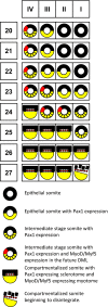
Schematic summary of the maturation stages of the caudalmost somites I–IV from HH‐stages 20–27.

Similar articles

Cited by

References

    1. Bellairs R, Sanders EJ (1986) Somitomeres in the chick tail bud: an SEM study. Anat Embryol (Berl) 175, 235–240. - PubMed
    1. Berti F, Nogueira JM, Wohrle S, et al. (2015) Time course and side‐by‐side analysis of mesodermal, pre‐myogenic, myogenic and differentiated cell markers in the chicken model for skeletal muscle formation. J Anat 227, 361–382. - PMC - PubMed
    1. Borman WH, Yorde DE (1994) Analysis of chick somite myogenesis by in situ confocal microscopy of desmin expression. J Histochem Cytochem 42, 265–272. - PubMed
    1. Borman WH, Urlakis KJ Jr, Yorde DE (1994) Analysis of the in vivo myogenic status of chick somites by desmin expression in vitro . Dev Dyn 199, 268–279. - PubMed
    1. Brand‐Saberi B, Ebensperger C, Wilting J, et al. (1993) The ventralizing effect of the notochord on somite differentiation in chick embryos. Anat Embryol (Berl) 188, 239–245. - PubMed

LinkOut - more resources