

Isolation and Culture of Skin-Derived Differentiated and Stem-Like Cells Obtained from the Arabian Camel (Camelus dromedarius)
- PMID: 31226810
- PMCID: PMC6616910
- DOI: 10.3390/ani9060378
Isolation and Culture of Skin-Derived Differentiated and Stem-Like Cells Obtained from the Arabian Camel (Camelus dromedarius)
Abstract
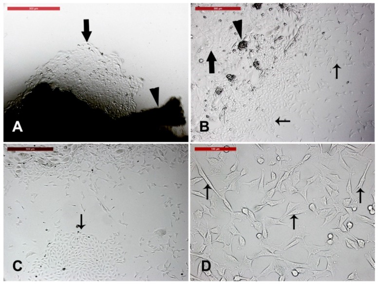
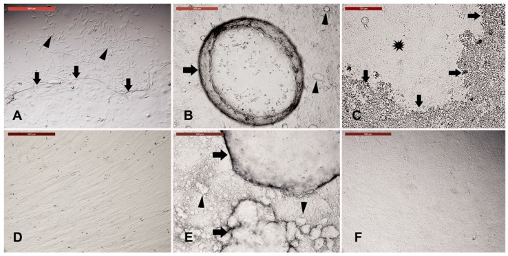
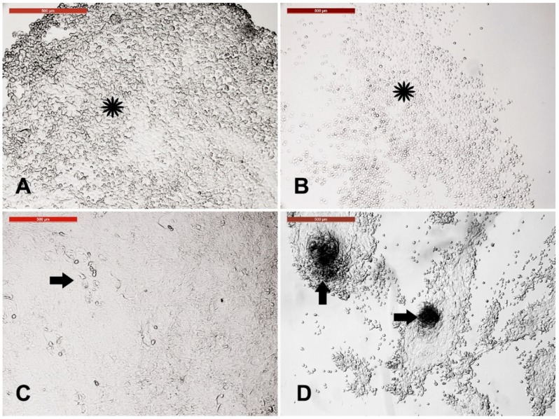
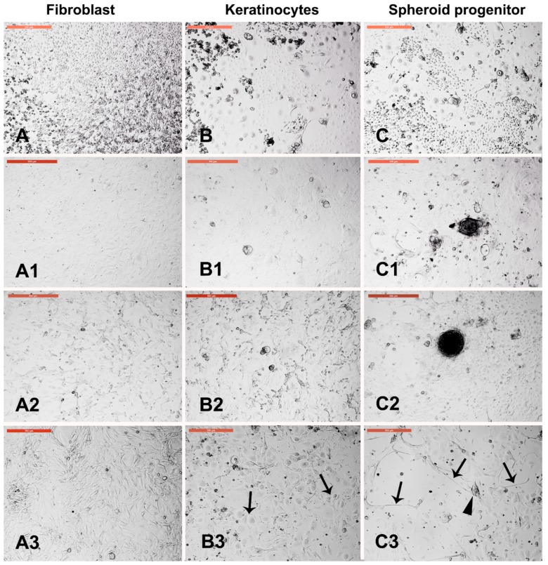
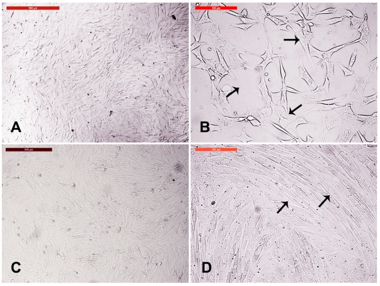
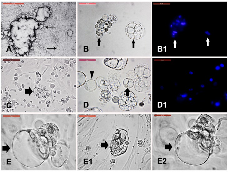
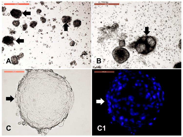
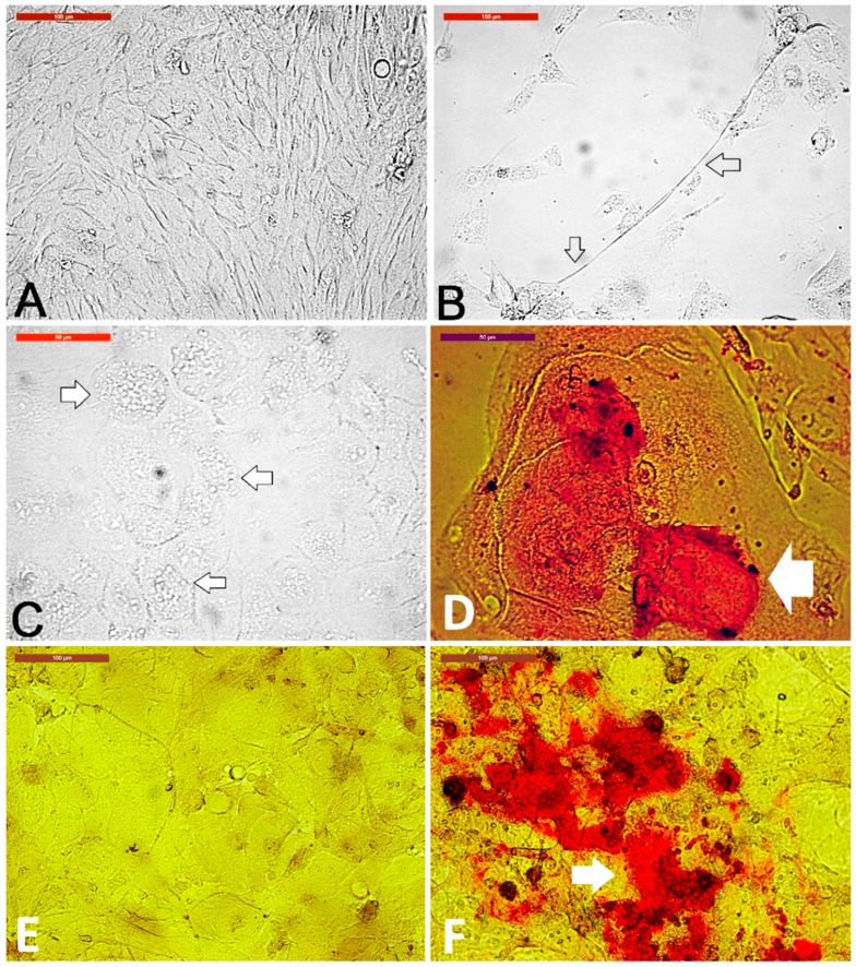
Elite camels often suffer from massive injuries. Thus, there is a pivotal need for a cheap and readily available regenerative medicine source. We isolated novel stem-like cells from camel skin and investigated their multipotency and resistance against various stresses. Skin samples were isolated from ears of five camels. Fibroblasts, keratinocytes, and spheroid progenitors were extracted. After separation of different cell lines by trypsinization, all cell lines were exposed to heat shock. Then, fibroblasts and dermal cyst-forming cells were examined under cryopreservation. Dermal cyst-forming cells were evaluated for resistance against osmotic pressure. The results revealed that resistance periods against trypsin were 1.5, 4, and 7 min for fibroblasts, keratinocytes, and spheroid progenitors, respectively. Furthermore, complete recovery of different cell lines after heat shock along with the differentiation of spheroid progenitors into neurons was observed. Fibroblasts and spheroid progenitors retained cell proliferation after cryopreservation. Dermal cyst-forming cells regained their normal structure after collapsing by osmotic pressure. The spheroid progenitors incubated in the adipogenic, osteogenic, and neurogenic media differentiated into adipocyte-, osteoblast-, and neuron-like cells, respectively. To the best of our knowledge, we isolated different unique cellular types and stem-like cells from the camel skin and examined their multipotency for the first time.
Keywords: camel; cell culture; differentiation; skin; stem cells.
Conflict of interest statement
The authors declare that they have no conflicts of interest.
Figures
Similar articles
-
Comparative Study of Biological Characteristics, and Osteoblast Differentiation of Mesenchymal Stem Cell Established from Camelus dromedarius Skeletal Muscle, Dermal Skin, and Adipose Tissues.Animals (Basel). 2021 Apr 4;11(4):1017. doi: 10.3390/ani11041017. Animals (Basel). 2021. PMID: 33916532 Free PMC article.
-
Isolation, characterization, and mesodermic differentiation of stem cells from adipose tissue of camel (Camelus dromedarius).In Vitro Cell Dev Biol Anim. 2013 Feb;49(2):147-54. doi: 10.1007/s11626-012-9578-9. Epub 2013 Jan 9. In Vitro Cell Dev Biol Anim. 2013. PMID: 23299319
-
Isolation and characterization of the trophectoderm from the Arabian camel (Camelus dromedarius).Placenta. 2017 Sep;57:113-122. doi: 10.1016/j.placenta.2017.06.015. Epub 2017 Jun 17. Placenta. 2017. PMID: 28863999
-
The current perspectives of dromedary camel stem cells research.Int J Vet Sci Med. 2018 Feb 14;6(Suppl):S27-S30. doi: 10.1016/j.ijvsm.2018.01.002. eCollection 2018. Int J Vet Sci Med. 2018. PMID: 30761317 Free PMC article. Review.
-
Cellular and Molecular Adaptation of Arabian Camel to Heat Stress.Front Genet. 2019 Jun 19;10:588. doi: 10.3389/fgene.2019.00588. eCollection 2019. Front Genet. 2019. PMID: 31275361 Free PMC article. Review.
Cited by
-
Isolation, characterization, and cryopreservation of collared peccary skin-derived fibroblast cell lines.PeerJ. 2020 Jun 3;8:e9136. doi: 10.7717/peerj.9136. eCollection 2020. PeerJ. 2020. PMID: 32547858 Free PMC article.
-
Industrial Research and Development on the Production Process and Quality of Cultured Meat Hold Significant Value: A Review.Food Sci Anim Resour. 2024 May;44(3):499-514. doi: 10.5851/kosfa.2024.e20. Epub 2024 May 1. Food Sci Anim Resour. 2024. PMID: 38765282 Free PMC article. Review.
-
Changes in Cell Vitality, Phenotype, and Function of Dromedary Camel Leukocytes After Whole Blood Exposure to Heat Stress in vitro.Front Vet Sci. 2021 Apr 9;8:647609. doi: 10.3389/fvets.2021.647609. eCollection 2021. Front Vet Sci. 2021. PMID: 33898545 Free PMC article.
-
Thermotolerance of camel (Camelus dromedarius) somatic cells affected by the cell type and the dissociation method.Environ Sci Pollut Res Int. 2019 Oct;26(28):29490-29496. doi: 10.1007/s11356-019-06208-5. Epub 2019 Aug 21. Environ Sci Pollut Res Int. 2019. PMID: 31435907
-
Comparative Study of Biological Characteristics, and Osteoblast Differentiation of Mesenchymal Stem Cell Established from Camelus dromedarius Skeletal Muscle, Dermal Skin, and Adipose Tissues.Animals (Basel). 2021 Apr 4;11(4):1017. doi: 10.3390/ani11041017. Animals (Basel). 2021. PMID: 33916532 Free PMC article.
References
-
- Gahlot T., Chouhan D. Fractures in dromedary (Camelus dromedarius)—A retrospective study. J. Camel Pract. Res. 1994;1:9–14.
-
- Sheyn D., Ben-David S., Shapiro G., De Mel S., Bez M., Ornelas L., Sahabian A., Sareen D., Da X., Pelled G. Human induced pluripotent stem cells differentiate into functional mesenchymal stem cells and repair bone defects. Stem Cells Transl. Med. 2016;5:1447–1460. doi: 10.5966/sctm.2015-0311. - DOI - PMC - PubMed
-
- Bhakat C., Sahani M. Scope of value addition to camel hide. Nat. Prod. Rad. 2005;4:387–390.
-
- Fowler M. Medicine and Surgery of Camelids. John Wiley & Sons; Hoboken, NJ, USA: 2011. Integumentary System; pp. 289–310.
Grants and funding
LinkOut - more resources
Full Text Sources
