OCT Angiography Biomarkers for Predicting Visual Outcomes after Ranibizumab Treatment for Diabetic Macular Edema
- PMID: 31227330
- PMCID: PMC6921516
- DOI: 10.1016/j.oret.2019.04.027
OCT Angiography Biomarkers for Predicting Visual Outcomes after Ranibizumab Treatment for Diabetic Macular Edema
Abstract
Purpose: To correlate quantitative OCT angiography (OCTA) biomarkers with clinical features and to predict the extent of visual improvement after ranibizumab treatment for diabetic macular edema (DME) with OCTA biomarkers.
Design: Retrospective, longitudinal study in Taiwan.
Participants: Fifty eyes of 50 patients with DME and 22 eyes of 22 healthy persons, with the exception of cataract and refractive error, from 1 hospital.
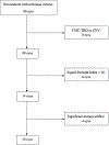
Methods: Each eye underwent OCT angiography (RTVue XR Avanti System with AngioVue software version 2017.1; Optovue, Fremont, CA), and 3×3-mm2 en face OCTA images of the superficial layer and the deep layer were obtained at baseline and after 3 monthly injections of ranibizumab in the study group. OCT angiography images also were acquired from the control group.
Main outcome measures: Five OCTA biomarkers, including foveal avascular zone (FAZ) area (FAZ-A), FAZ contour irregularity (FAZ-CI), average vessel caliber (AVC), vessel tortuosity (VT), and vessel density (VD), were analyzed comprehensively. Best-corrected visual acuity (BCVA) and central retinal thickness (CRT) also were obtained. Student t tests were used to compare the OCTA biomarkers between the study group and the control group. Linear regression models were used to evaluate the correlations between the baseline OCTA biomarkers and the changes of BCVA and CRT after treatment.
Results: Eyes with DME had larger AVC, VT, FAZ-A, and FAZ-CI and lower VD than those in the control group (P < 0.001 for all). After the loading ranibizumab treatment, these OCTA biomarkers improved but did not return to normal levels. Among all biomarkers, higher inner parafoveal VD in the superficial layer at baseline correlated most significantly with visual gain after treatment in the multiple regression model with adjustment for CRT and ellipsoid zone disruption (P < 0.001). To predict visual improvement, outer parafoveal VD in the superficial layer at the baseline showed the largest area under the receiver operating characteristic curve (0.787; P = 0.004). No baseline OCTA biomarkers showed any significant correlation specifically with anatomic improvement.
Conclusions: For eyes with DME, parafoveal VD in the superficial layer at baseline was an independent predictor for visual improvement after the loading ranibizumab treatment.
Copyright © 2019 American Academy of Ophthalmology. Published by Elsevier Inc. All rights reserved.
Figures


References
-
- Wong TY, Sun J, Kawasaki R, et al. Guidelines on diabetic eye care: the International Council of Ophthalmology recommendations for screening, follow-up, referral, and treatment based on resource settings. Ophthalmology. 2018;125: 1608–1622. - PubMed
-
- Lattanzio R, Cicinelli MV, Bandello F. Intravitreal steroids in diabetic macular edema. Dev Ophthalmol. 2017;60:78–90. - PubMed
-
- Douvali M, Chatziralli IP, Theodossiadis PG, et al. Effect of macular ischemia on intravitreal ranibizumab treatment for diabetic macular edema. Ophthalmologica. 2014;232: 136–143. - PubMed
-
- Serizawa S, Ohkoshi K, Minowa Y, Soejima K. Interdigitation zone band restoration after treatment of diabetic macular edema. Curr Eye Res. 2016;41:1229–1234. - PubMed
Publication types
MeSH terms
Substances
Grants and funding
LinkOut - more resources
Full Text Sources
Medical
Research Materials

