. 2019 Jul 9;116(28):14309-14318.
doi: 10.1073/pnas.1900774116. Epub 2019 Jun 21.

Cryo-EM structure of OSCA1.2 from Oryza sativa elucidates the mechanical basis of potential membrane hyperosmolality gating

Affiliations

Cryo-EM structure of OSCA1.2 from Oryza sativa elucidates the mechanical basis of potential membrane hyperosmolality gating

Koustav Maity et al. Proc Natl Acad Sci U S A. .

Abstract

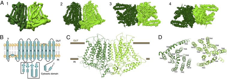
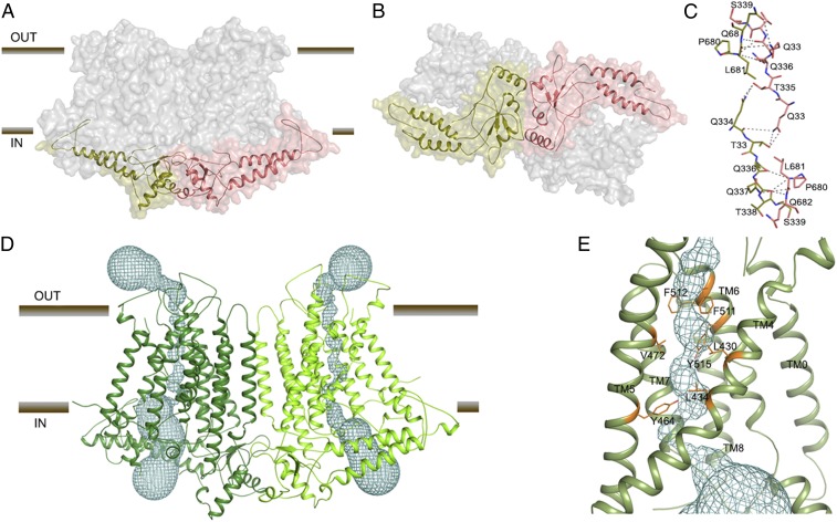
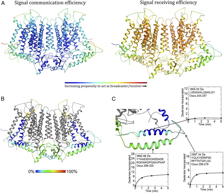
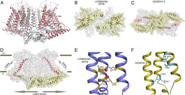
Sensing and responding to environmental water deficiency and osmotic stresses are essential for the growth, development, and survival of plants. Recently, an osmolality-sensing ion channel called OSCA1 was discovered that functions in sensing hyperosmolality in Arabidopsis Here, we report the cryo-electron microscopy (cryo-EM) structure and function of an OSCA1 homolog from rice (Oryza sativa; OsOSCA1.2), leading to a model of how it could mediate hyperosmolality sensing and transport pathway gating. The structure reveals a dimer; the molecular architecture of each subunit consists of 11 transmembrane (TM) helices and a cytosolic soluble domain that has homology to RNA recognition proteins. The TM domain is structurally related to the TMEM16 family of calcium-dependent ion channels and lipid scramblases. The cytosolic soluble domain possesses a distinct structural feature in the form of extended intracellular helical arms that are parallel to the plasma membrane. These helical arms are well positioned to potentially sense lateral tension on the inner leaflet of the lipid bilayer caused by changes in turgor pressure. Computational dynamic analysis suggests how this domain couples to the TM portion of the molecule to open a transport pathway. Hydrogen/deuterium exchange mass spectrometry (HDXMS) experimentally confirms the conformational dynamics of these coupled domains. These studies provide a framework to understand the structural basis of proposed hyperosmolality sensing in a staple crop plant, extend our knowledge of the anoctamin superfamily important for plants and fungi, and provide a structural mechanism for potentially translating membrane stress to transport regulation.

Keywords: channel; cryo-EM; osmotic stress; rice; structure.


Conflict of interest statement

The authors declare no conflict of interest.

Figures

Fig. 1.
Fig. 1.
Cryo-EM structure of the OsOSCA1.2 protein. (A, Left to Right) (1) Parallel to membrane plane view of unsharpened cryo-EM density map used for initial chain tracing and (2–4) sharpened 4.9-Å map used for model building and refinement [membrane plane view (2), extracellular view (3), and intracellular view (4)]. (B) Protein topology of OsOSCA1.2. The OsOSCA1.2 model is shown in the TM plane (C) and from the extracellular side (D).
Fig. 2.
Fig. 2.
OsOSCA1.2 dimer interface and transport pathway. (A) OsOSCA1.2 surface representation. The TM domain is shown in gray, and the cytoplasmic domain is colored red and green. (B) View of OsOSCA1.2 from the cytoplasmic side. (C) Dimer interface residues. (D) Location of the predicted transport pathway in both subunits of OsOSCA1.2. The transport pathway is depicted as a cyan mesh. (E) Close-up view of the neck region, showing the residues “gating” the transport pathway.
Fig. 3.
Fig. 3.
Computational and experimental dynamics of OsOSCA1.2. (A) Results of OsOSCA1.2 embedded in the membrane using the DynOmics suite. Panels show a color-coded map superimposed on the model illustrating signal communication (Left) and receiving (Right) efficiency. Regions that are colored red are more active, while those that are colored blue are inactive with regard to molecular dynamics prediction. (B) Relative uptake after 5 min of exchange. The structure is color-scaled and superimposed on the model. Regions colored gray yielded no detectable peptide fragments. (C) Close-up view of the extended and gating helix. Uptake plots for selected peptides are shown. Corresponding protein segments are outlined.
Fig. 4.
Fig. 4.
Structural comparisons of OsOSCA1.2 with other TMEM and OSCA structures. (A) Superposition of OsOSCA1.2 and AtOSCA1 (Protein Data Bank ID code 5YD1; ref. 8). TM1, TM2, TM9, and TM10 (shown in gray) close to the lipid-filled cleft are nearly superimposable and have little relative movement. Transport pathway-lining helices (TM3–TM7) showed significant movement, along with TM0 and TM8 (shown in red). (B) mTMEM16A soluble domains from the intercellular side are separated. (C) OsOSCA1.2 intracellular soluble domains are together and communicate with the gating helix TM6. (D) General mechanism of OsOSCA1.2 shown in the plane of the lipid membrane. Lateral tension on the inner leaflet side of the lipid bilayer causes a conformational change in the extended helices of the soluble domain, which is coupled to the gating helix TM6 opening of the transport pathway. (E) Calcium binding site residues of Ca2+-activated chloride channel mTMEM16A. Calcium ions are shown as red spheres. (F) Corresponding region of OsOSCA1.2 with charged and polar residues shown in cyan.

References

    1. Yuan F., et al. , OSCA1 mediates osmotic-stress-evoked Ca2+ increases vital for osmosensing in Arabidopsis. Nature 514, 367–371 (2014). - PubMed
    1. Knight H., Trewavas A. J., Knight M. R., Calcium signalling in Arabidopsis thaliana responding to drought and salinity. Plant J. 12, 1067–1078 (1997). - PubMed
    1. Stephan A. B., Kunz H. H., Yang E., Schroeder J. I., Rapid hyperosmotic-induced Ca2+ responses in Arabidopsis thaliana exhibit sensory potentiation and involvement of plastidial KEA transporters. Proc. Natl. Acad. Sci. U.S.A. 113, E5242–E5249 (2016). - PMC - PubMed
    1. Choi W. G., Toyota M., Kim S. H., Hilleary R., Gilroy S., Salt stress-induced Ca2+ waves are associated with rapid, long-distance root-to-shoot signaling in plants. Proc. Natl. Acad. Sci. U.S.A. 111, 6497–6502 (2014). - PMC - PubMed
    1. Murthy S. E., et al. , OSCA/TMEM63 are an evolutionarily conserved family of mechanically activated ion channels. eLife 7, e41844 (2018). - PMC - PubMed

Publication types

MeSH terms

Associated data