. 2019 Jun 21;10(1):2746.
doi: 10.1038/s41467-019-10723-x.

α7 nicotinic acetylcholine receptor upregulation by anti-apoptotic Bcl-2 proteins

Affiliations

α7 nicotinic acetylcholine receptor upregulation by anti-apoptotic Bcl-2 proteins

G Brent Dawe et al. Nat Commun. .

Abstract

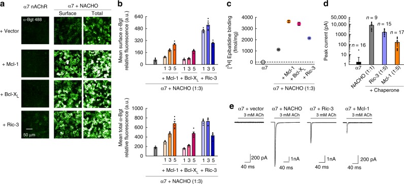
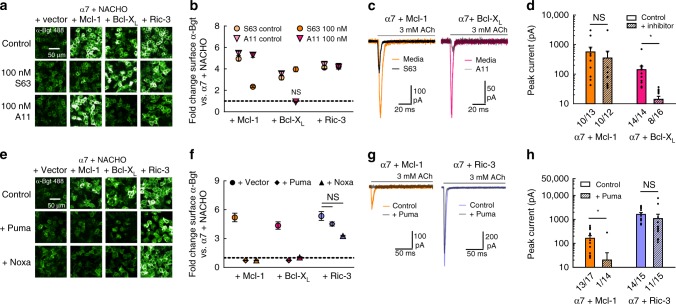
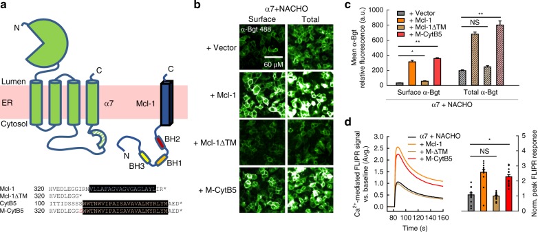
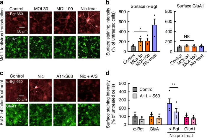
Nicotinic acetylcholine receptors (nAChRs) mediate and modulate synaptic transmission throughout the brain, and contribute to learning, memory, and behavior. Dysregulation of α7-type nAChRs in neuropsychiatric as well as immunological and oncological diseases makes them attractive targets for pharmaceutical development. Recently, we identified NACHO as an essential chaperone for α7 nAChRs. Leveraging the robust recombinant expression of α7 nAChRs with NACHO, we utilized genome-wide cDNA library screening and discovered that several anti-apoptotic Bcl-2 family proteins further upregulate receptor assembly and cell surface expression. These effects are mediated by an intracellular motif on α7 that resembles the BH3 binding domain of pro-apoptotic Bcl-2 proteins, and can be blocked by BH3 mimetic Bcl-2 inhibitors. Overexpression of Bcl-2 member Mcl-1 in neurons enhanced surface expression of endogenous α7 nAChRs, while a combination of chemotherapeutic Bcl2-inhibitors suppressed neuronal α7 receptor assembly. These results demonstrate that Bcl-2 proteins link α7 nAChR assembly to cell survival pathways.


Conflict of interest statement

The authors declare no competing interests except that authors are all full-time employees of Janssen Pharmaceutical Companies of Johnson and Johnson.

Figures

Fig. 1
Fig. 1
Bcl-2 proteins enhance assembly and functional expression of α7 nAChRs. a Immunofluorescent staining of HEK293T cells cotransfected with cDNAs encoding α7, or α7 and NACHO, along with Mcl-1, Bcl-XL, or Ric-3 (at 1:3:5 cDNA ratio, respectively). Surface and total staining were performed before and after membrane permeabilization, respectively, using α-Bgt conjugated to a fluorescent probe. b Quantification of specific α-Bgt labeling, expressed as fluorescence intensity (n = 5). cDNA ratios are indicated relative to the α7-encoding vector. At 5× cDNA ratios, the increase in surface (top) and total (bottom) α-Bgt labeling produced by Mcl-1, Bcl-XL, or Ric-3 cotransfection was significant (p < 0.001). The same cDNA combinations were transfected at different passage numbers and yielded similar results. c Binding of 10 nM [3H]epibatidine to HEK293T cell membranes cotransfected with α7, NACHO, and other proteins as indicated (at 1:3:5 cDNA ratio, respectively), with measurements taken from multiple samples during a single experiment (n = 6). The increase in binding produced by Mcl-1, Bcl-XL, or Ric-3 cotransfection atop α7 and NACHO was significant (p < 1e−5). d, e Summary graph of agonist-evoked peak currents for HEK293T cells cotransfected with specified cDNAs along with eGFP, with n values listed for each condition. Versus α7 expressed alone, where only 1 of 16 cells responded to ACh, coexpression of either NACHO, Ric-3, or Mcl-1 yielded significant effects (p < 0.01). Ratios of each cDNA are indicated relative to the α7-encoding vector. Representative whole-cell current responses elicited from the conditions in panel (d) are also shown (e). All data are means ± SEM; p values from two-sample t test
Fig. 2
Fig. 2
Chemical inhibitors and BH3-only proteins eliminate Bcl-2-mediated upregulation of α7 nAChRs. a Fluorescent α-Bgt labeling of nonpermeabilized HEK293T cells cotransfected with cDNAs encoding α7 and NACHO, along with other proteins indicated, at a 1:3:4 respective ratio. b Surface α-Bgt labeling with S63 or A11 inhibitors, presented as a fold change in fluorescence intensity (n = 5 treated, n = 10 control), relative to α7 and NACHO expressed alone. Enhancement of fluorescence remained significant in all cases (p < 0.01) except cells expressing Bcl-XL treated with 100 nM A11 (p = 0.13). c, d Representative whole-cell current responses elicited from HEK293T cells expressing α7, and either Mcl-1 or Bcl-XL (1:5 cDNA ratio), with or without 24 h incubation at 30 °C in the inhibitors A11 and S63 (1 µM), respectively (c). Summary graph of agonist-evoked peak currents (*p < 0.01) with the number of responsive cells indicated (d). The reduction in peak response was significant for A11, but not S63 (p = 0.56) treatment. e Fluorescent α-Bgt labeling of nonpermeabilized HEK293T cells cotransfected with cDNAs encoding α7 and NACHO, along with other proteins indicated, including Puma or Noxa at a 1:3:4:4 (α7: NACHO: other: BH3) cDNA ratio. f Fold change in fluorescence intensity for α-Bgt labeled cells in the presence of BH3 only proteins (n = 5), relative to α7 and NACHO expressed alone. Coexpression of Puma or Noxa significantly reduced fluorescence intensity in the presence of Mcl-1 or Bcl-XL (p < 0.01), but not Ric-3 (p ≥ 0.22). g, h Representative whole-cell current responses elicited from HEK293T cells cotransfected with cDNAs encoding α7 and either Mcl-1 or Ric-3, ±Puma at a 1:5:5 respective cDNA ratio (g). Summary graph of agonist-evoked peak currents (*p < 0.01) with the number of responsive cells indicated (h). Control values taken from Fig. 1d. The reduction in peak response was significant for Mcl-1, but not Ric-3 (p = 0.41) expressing cells. All data are means ± SEM; p values from two-sample t test. For all α-Bgt labeling experiments, the same cDNA transfection conditions were repeated at different passage numbers and yielded similar results
Fig. 3
Fig. 3
ER-anchoring of the Mcl-1 TM segment is critical for regulation of α7 nAChR assembly. a Topological illustration of the α7 nAChR subunit and Mcl-1 within the ER membrane (top), and amino acid sequence alignment of various Mcl-1 constructs examined with transmembrane regions highlighted in black (bottom). b Fluorescent α-Bgt labeling of nonpermeabilized and permeabilized HEK293T cells cotransfected with cDNAs encoding α7 and NACHO, along with empty vector, wild-type Mcl-1, a truncated Mcl-1 construct lacking the C-terminal TM segment (Mcl-1ΔTM), or a chimeric Mcl-1 containing the TM sequence of cytochrome B5 in place of its own (M-CytB5) at a 1:3:4 respective ratio. c Quantification of fluorescence intensity from surface and total labeling of α7 nAChRs by α-Bgt when coexpressed with NACHO and various Mcl-1 constructs (n = 5, *p < 0.01, **p < 0.001). Increases in total α-Bgt labeling were significant (α = 0.017) for cells expressing Mcl-1 and M-CytB5, but not Mcl-1ΔTM (p = 0.04), atop α7 and NACHO. The same cDNA combinations were transfected at different passage numbers and yielded similar results. d Averaged FLIPR traces showing 100 µM nicotine-evoked Ca2+ flux through HEK293T cells cotransfected with cDNAs encoding the α7 nAChR, NACHO, and various Mcl-1 constructs at a 1:1:5 respective ratio (left). Also shown is the mean peak response above baseline (n = 15, *p < 1e−5), normalized to the background fluorescence signal (right). Increases in FLIPR response were significant for cells expressing Mcl-1 and M-CytB5, but not Mcl-1ΔTM (p = 0.60), atop α7 and NACHO. Data shown are from an individual experiment that was replicated. All data are means ± SEM; p values from two-sample t test
Fig. 4
Fig. 4
Mutations in a BH3-like motif of α7 nAChRs attenuate Bcl-2-mediated upregulation. a Cartoon illustration of α7 nAChR topology, including the region removed in the del 347 mutant and location of the pre-M4 helix (left). Also shown is a sequence alignment between the pre-M4 helix and BH3 domains of several Bcl-2 family proteins (right), color-coded by hydrophobicity, where hydrophobic (green), acidic (red), and basic (blue) residues at key positions are indicated. b Structure of Bcl-XL bound by the BH3 segment of Bak (PDB: 1BXL) with hydrophobic residues on Bak mediating the interaction highlighted (teal). c Fluorescent α-Bgt labeling of permeabilized HEK293T cells cotransfected with cDNAs encoding wild-type or mutant α7 and NACHO, along with other proteins, at a 1:3:4 respective ratio. d Quantification of fluorescence intensity from α7 mutants labeled by α-Bgt (top) and the fold change in fluorescence intensity relative to α7 and NACHO expressed alone (bottom; n = 5). The fold effect of Mcl-1 and Bcl-XL coexpression was significantly reduced when comparing wild-type with mutant α7 subunits (p < 0.01), though Ric-3-mediated upregulation did not differ significantly for any mutant except del 347 (which was further enhanced). The same cDNA combinations were transfected at different passage numbers and yielded similar results. e Representative whole-cell current responses elicited from HEK293T cells transfected with cDNAs encoding wild-type or mutant α7 receptors, alone or with Mcl-1, or Ric-3 at a 1:5 (α7: other) cDNA ratio. Cells were incubated 24 h at 30 °C prior to recording. f Summary graph of agonist-evoked peak currents for transfections described in panel (e) (*p < 0.01) with the n value for each condition indicated. Values for wild-type α7 with Mcl-1 and Bcl-XL taken from data set in Fig. 2d. For the wild-type receptor, but not the I436A mutant (p = 0.03, WT; p = 0.22, I436A; two-sample one-tail t test), Mcl-1 significantly increased peak currents. All data are means ± SEM; p values from two-sample t test
Fig. 5
Fig. 5
Bcl-2 proteins regulate α7 nAChRs in hippocampal neurons. a Fluorescent labeling of nonpermeabilized rat hippocampal neurons (DIV 20) transduced with lentivirus encoding human Mcl-1 at DIV7 or incubated with 100 µM nicotine (Nic) since DIV 15. Representative images of surface α-Bgt and surface GluA1 (AMPA receptor) labeling of neurons are shown. b Quantification of surface α-Bgt (left) and surface GluA1 (right) labeling, expressed as a percentage of the intensity from untreated neurons. Two multiplicities of infection (MOI 30 and 100) were compared to ensure maximal lentiviral transduction occurred. Data are averaged from three experiments on different cultures (*p < 0.05). At MOI 100, there was a significant increase in α-Bgt staining, but not GluA1 staining (p = 0.41, paired one-tail t test). c Fluorescent labeling of nonpermeabilized rat hippocampal neurons (DIV 21) treated for 96 h with the Bcl-XL inhibitor A-1155463 (A11, 10 nM) and Mcl-1 inhibitor S-63845 (S63, 10 nM), with or without coincubation in 100 µM nicotine (Nic) to promote α7 nAChR surface expression. Representative images of surface α-Bgt and surface GluA1 labeling are shown. d Quantification of surface α-Bgt and surface GluA1 fluorescence labeling intensity, expressed as a percentage of the fluorescence from untreated neurons. Data are averaged from four experiments on different cultures (**p < 0.01). With nicotine pretreatment, Bcl-2 inhibitors induced a significant decrease in α-Bgt staining, but not GluA1 staining (p = 0.09, paired one-tail t test). All data are means ± SEM

Similar articles

Cited by

References

    1. Albuquerque EX, Pereira EF, Alkondon M, Rogers SW. Mammalian nicotinic acetylcholine receptors: from structure to function. Physiol. Rev. 2009;89:73–120. doi: 10.1152/physrev.00015.2008. - DOI - PMC - PubMed
    1. Le Novere N, Corringer PJ, Changeux JP. The diversity of subunit composition in nAChRs: evolutionary origins, physiologic and pharmacologic consequences. J. Neurobiol. 2002;53:447–456. doi: 10.1002/neu.10153. - DOI - PubMed
    1. Gotti C, Zoli M, Clementi F. Brain nicotinic acetylcholine receptors: native subtypes and their relevance. Trends Pharm. Sci. 2006;27:482–491. doi: 10.1016/j.tips.2006.07.004. - DOI - PubMed
    1. Couturier S, et al. A neuronal nicotinic acetylcholine receptor subunit (alpha 7) is developmentally regulated and forms a homo-oligomeric channel blocked by alpha-BTX. Neuron. 1990;5:847–856. doi: 10.1016/0896-6273(90)90344-F. - DOI - PubMed
    1. Li SX, et al. Ligand-binding domain of an alpha7-nicotinic receptor chimera and its complex with agonist. Nat. Neurosci. 2011;14:1253–1259. doi: 10.1038/nn.2908. - DOI - PMC - PubMed

MeSH terms