. 2019 Jun 21;9(1):8998.
doi: 10.1038/s41598-019-44534-3.

Phrixotrix luciferase and 6'-aminoluciferins reveal a larger luciferin phenolate binding site and provide novel far-red combinations for bioimaging purposes

Affiliations

Phrixotrix luciferase and 6'-aminoluciferins reveal a larger luciferin phenolate binding site and provide novel far-red combinations for bioimaging purposes

V R Bevilaqua et al. Sci Rep. .

Abstract

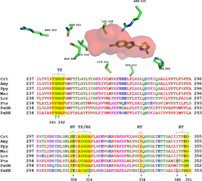
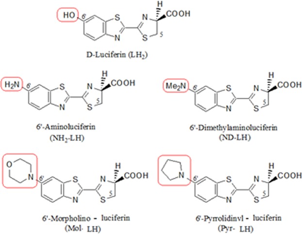
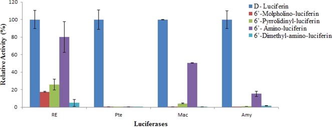
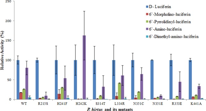
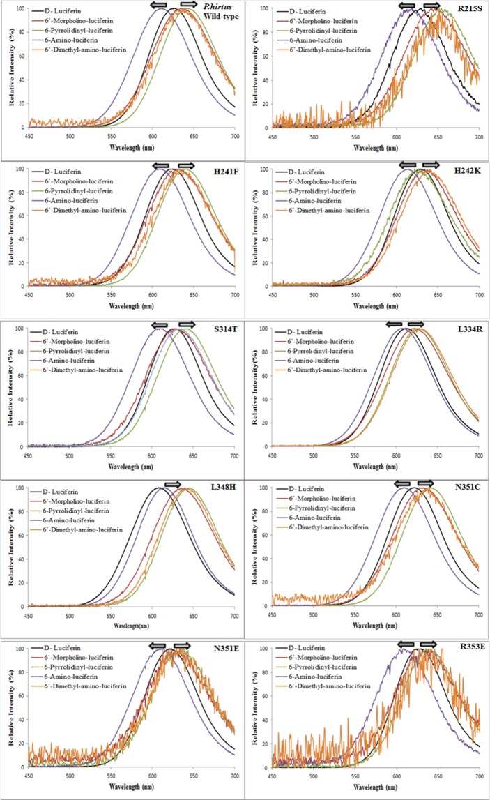
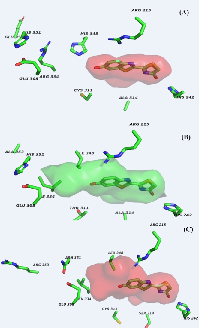
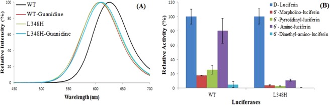
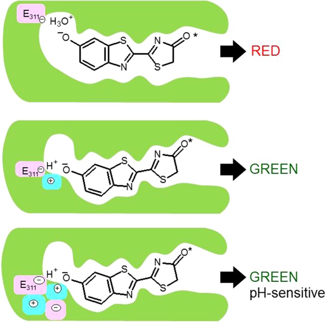
How the unique luciferase of Phrixothrix hirtus (PxRE) railroad worm catalyzes the emission of red bioluminescence using the same luciferin of fireflies, remains a mystery. Although PxRE luciferase is a very attractive tool for bioanalysis and bioimaging in hemoglobin rich tissues, it displays lower quantum yield (15%) when compared to green emitting luciferases (>40%). To identify which parts of PxRE luciferin binding site (LBS) determine bioluminescence color, and to develop brighter and more red-shifted emitting luciferases, we compared the effects of site-directed mutagenesis and of larger 6'-substituted aminoluciferin analogues (6'-morpholino- and 6'-pyrrolidinyl-LH) on the bioluminescence properties of PxRE and green-yellow emitting beetle luciferases. The effects of mutations in the benzothiazolyl and thiazolyl parts of PxRE LBS on the KM and catalytic efficiencies, indicated their importance for luciferin binding and catalysis. However, the absence of effects on the bioluminescence spectrum indicated a less interactive LBS in PxRE during light emission. Mutations at the bottom of LBS of PxRE blue-shifted the spectra and increased catalytic efficiency, suggesting that lack of interactions of this part of LBS with excited oxyluciferin phenolate underlie red light emission. The much higher bioluminescence activity and red-shifted spectra of PxRE luciferase with 6'-morpholino- (634 nm) and 6'-pyrrolidinyl-luciferins (644 nm), when compared to other beetle luciferases, revealed a larger luciferin phenolate binding pocket. The size and orientation of the side-chains of L/I/H348 are critical for amino-analogues accommodation and modulate bioluminescence color, affecting the interactions and mobility of excited oxyluciferin phenolate. The PxRE luciferase and 6'-aminoluciferins provide potential far-red combinations for bioimaging applications.


Conflict of interest statement

The authors declare no competing interests.

Figures

Figure 1
Figure 1
(Upper) Phrixotrix hirtus railroad worm and (lower) Red and far-red bioluminescence of E. coli expressing PxRE luciferase in presence of (A) D-luciferin; (B) 6'-morpholinoluciferin and (C) 6'-pyrrolidinylluciferin.
Figure 2
Figure 2
(Upper panel) threedimensional model of PxRE luciferase showing the location of the investigated residues; (Lower panel) multialignment of the luciferin binding site (LBS) segments (yellow shadow) of Phrixotrix hirtus red emitting luciferase with other beetle luciferases in the region between residues 238–358 (Photinus pyralis sequence): (BZ) Benzothiazole; (TZ) Thiazole; (BZ/TZ) Benzothiazole/Thiazole; (BT) Bottom; (red) hydrophobic residues; (green) polar residues; (blue) negatively charged residues; (pink) positively charged residue; (gray shadow) investigated residues; (Pte) Pyrearinus termitilluminans larval click beetle; (Mac) Macrolampis sp2 firefly; (Ppy) Photinus pyralis firefly; (Crt) Cratomorphus distinctus firefly; (Amy) Amydetes vivianii firefly; (Rol) Ragophtalmus ohbai starworm; (PxGR) Phrixotrix vivianii railroadworm green emitting and (PxRE) Phrixotrix hirtus railroad worm red emitting luciferase.
Figure 3
Figure 3
Structures of D-luciferin, 6′-aminoluciferin, and 6′-substituted amino luciferin analogues. The portions in red show the 6′-amino and substituted 6′-amino groups.
Figure 4
Figure 4
Relative overall and oxidative activities of P. hirtus red-emitting luciferase and its mutants: (red) overall bioluminescent activity starting with ATP and luciferin; (Blue) oxidative bioluminescent activity starting with luciferyl-adenylate.
Figure 5
Figure 5
Relative activity of beetle luciferases with firefly D-luciferin and 6′-amino-analogues: Macrolampis sp2 and Amydetes vivianii firefly, P. termitilluminans click beetle and P. hirtus railroadworm. The activities of each luciferases were normalized for D-luciferin.
Figure 6
Figure 6
Relative activity of P. hirtus red-emitting luciferase and mutants with 6′-amino-analogues and luciferin. The activities of each mutant luciferase were normalized for D-luciferin.
Figure 7
Figure 7
Bioluminescence spectra of beetle luciferases with 6′-amino-analogues.
Figure 8
Figure 8
Bioluminescence spectra of P. hirtus red emitting luciferase and its mutants with LH2, NH2-LH, and 6′-substituted amino analogues.
Figure 9
Figure 9
Active site modelling of beetle luciferases showing a larger phenol binding cavity in Phrixotrix hirtus red emitting luciferase: (A) Pyrearinus termitilluminans luciferase; (B) Phrixotrix vivianii luciferase; (C) P. hirtus red-emitting luciferase.
Figure 10
Figure 10
(Left panel) Bioluminescence spectra of Phrixotrix red-emitting luciferase mutant L348H in the presence and absence of guanidine; (Right panel) Bioluminescence activity with LH2, NH2-LH, and 6′-substituted amino analogues. The activities of each mutant luciferase were normalized for D-luciferin.
Figure 11
Figure 11
Proposed mechanism of bioluminescence color modulation by the active site of PxRE and green-emitting luciferases.

References

    1. Wood KV. The chemical mechanism and evolutionary development of beetle bioluminescence. Photochem. Photobiol. Sci. 1995;62:662–673. doi: 10.1111/j.1751-1097.1995.tb08714.x. - DOI
    1. Viviani VR. The origin, diversity and structure function relationships of insect luciferases. Cell. Mol. Life Sci. 2002;59:1833–1850. doi: 10.1007/PL00012509. - DOI - PMC - PubMed
    1. Roda A, Guardigli M, Michelini E, Mirasoli M. Bioluminescence in analytical chemistry and in vivo imaging. Trends Anal. Chem. 2009;28:307–322. doi: 10.1016/j.trac.2008.11.015. - DOI
    1. Asai, T., Nishi, S., Nakajima, Y. & Ohmiya, Y. Beetle luciferases in measuring gene expression and imaging cell functions in Luciferases and Fluorescent Proteins: Principles and Advances in Biotechnology and Bioimaging (eds Viviani, V. R. & Ohmiya, Y.) 151–160 (Transworld Research Network, 2007).
    1. Ugarova NN, Brovko LY. Protein structure and bioluminescence spectra for firefly bioluminescence. Luminescence. 2002;17:321–330. doi: 10.1002/bio.688. - DOI - PubMed

Publication types

MeSH terms