. 2019 Jun 21;9(1):9018.
doi: 10.1038/s41598-019-44957-y.

Molecular phenotyping of the surfaceome of migratory chondroprogenitors and mesenchymal stem cells using biotinylation, glycocapture and quantitative LC-MS/MS proteomic analysis

Affiliations

Molecular phenotyping of the surfaceome of migratory chondroprogenitors and mesenchymal stem cells using biotinylation, glycocapture and quantitative LC-MS/MS proteomic analysis

Csaba Matta et al. Sci Rep. .

Abstract

The complement of cell surface proteins, collectively referred to as the surfaceome, is a useful indicator of normal differentiation processes, and the development of pathologies such as osteoarthritis (OA). We employed biochemical and proteomic tools to explore the surfaceome and to define biomarkers in chondrogenic progenitor cells (CPC) derived from human OA knee articular cartilage. These cells have great therapeutic potential, but their unexplored biology limits their clinical application. We performed biotinylation combined with glycocapture and high throughput shotgun proteomics to define the surface proteome of human bone marrow mesenchymal stem cells (MSCs) and human CPCs. We prepared cell surface protein-enriched fractions from MSCs and CPCs, and then a proteomic approach was used to compare and evaluate protein changes between undifferentiated MSCs and CPCs. 1256 proteins were identified in the study, of which 791 (63%) were plasma membrane, cell surface or extracellular matrix proteins. Proteins constituting the surfaceome were annotated and categorized. Our results provide, for the first time, a repository of quantitative proteomic data on the surfaceome of two closely related cell types relevant to cartilage biology and OA. These results may provide novel insights into the transformation of the surfaceome during chondrogenic differentiation and phenotypic changes during OA development.


Conflict of interest statement

The authors declare no competing interests.

Figures
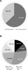
Figure 1
Figure 1
(A) Subcellular distribution of the surfaceome proteins identified following glycocapture. The surfaceome comprises the subproteome with proteins annotated as “plasma membrane”, “cell surface”, “cell membrane”, “extracellular” or “secreted”, as well as their associated proteins. (B) Surface proteins were further classified according to their presence/absence and relative expression in CPC and MSC, based on normalised quantitative data (cut-off: ±1.5 FC). Numbers in pie charts represent the actual numbers and relative percentages of proteins identified in each subgroup using all data from the PEAKS Studio protein identification export.
Figure 2
Figure 2
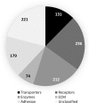
Classification of the identified 791 surface proteins in both CPCs and MSCs (combined). Surface proteins were categorised according to their GO annotations in the UniProt database (GO molecular function and/or GO biological process) into the following major functional groups: transporters, receptors, enzymes, extracellular matrix components, proteins involved in cell adhesion and cell junctions, and unclassified proteins. Note that some proteins appear in more than one category. Numbers in the pie chart represent the actual numbers of proteins classified into each subgroup.
Figure 3
Figure 3
Pie charts showing the differential expression of proteins in CPCs and MSCs in all 6 functional protein groups (cut-off: ±1.5 FC). Numbers in the pie charts represent the relative percentages of proteins in each subgroup using all data from the PEAKS Studio protein identification export.
Figure 4
Figure 4
(A) Protein profile heatmap generated from PEAKS Studio quantitation module, showing significantly differentially expressed proteins following quantitative LC-MS/MS analysis (cut-off fold change >1.5) utilising the Top 3 peptides from each protein. Cell colour represents the log2(ratio) to the average area across different samples. (B) Functional classification of significantly differentially expressed surface proteins. Surface proteins were categorised according to their GO annotations in the UniProt database into the major functional groups. Numbers in the pie chart represent the actual numbers and percentages of proteins classified into each subgroup.
Figure 5
Figure 5
Validation of the efficacy of the aminooxy-biotin based glycocapture method. (A) Western blot experiment performed on total cell lysates from human CPC and MSC cultures. After SDS–PAGE, protein bands were visualised using the Odyssey FC imaging system (Li-Cor) and bands were detected using the 800 nm (for KCNMA1) and 700 nm (for β-actin) channels. Optical density values of detected bands were calculated using the Image Studio software and the values for KCNMA1 were normalised to those of β-actin and then to MSC. Representative gel image. Full-length uncropped blots are presented in Fig. S2. Error bars are ± SD, n = 3. * indicates statistical significance (P < 0.05). (B) Intracellular distribution of KCNMA1 in MSCs and CPCs detected by immunocytochemistry. Primary antibodies were visualised using Alexa488-conjugated anti-rabbit secondary antibodies. Nuclear DNA was stained with DAPI. Data shown are representative out of 3 independent experiments. Scale bar, 50 µm.
Figure 6
Figure 6
Schematic overview of the experimental design used in this study.

References

    1. Cordwell SJ, Thingholm TE. Technologies for plasma membrane proteomics. Proteomics. 2010;10:611–627. doi: 10.1002/pmic.200900521. - DOI - PubMed
    1. Josic D, Clifton JG, Kovac S, Hixson DC. Membrane proteins as diagnostic biomarkers and targets for new therapies. Current opinion in molecular therapeutics. 2008;10:116–123. - PubMed
    1. Bausch-Fluck D, et al. The in silico human surfaceome. Proc Natl Acad Sci USA. 2018;115:E10988–E10997. doi: 10.1073/pnas.1808790115. - DOI - PMC - PubMed
    1. Hormann K, et al. A Surface Biotinylation Strategy for Reproducible Plasma Membrane Protein Purification and Tracking of Genetic and Drug-Induced Alterations. Journal of proteome research. 2016;15:647–658. doi: 10.1021/acs.jproteome.5b01066. - DOI - PubMed
    1. Weekes MP, et al. Comparative analysis of techniques to purify plasma membrane proteins. Journal of biomolecular techniques: JBT. 2010;21:108–115. - PMC - PubMed

Publication types

MeSH terms