. 2019 Oct:168:71-81.
doi: 10.1016/j.bcp.2019.06.017. Epub 2019 Jun 19.

Enterobactin, an iron chelating bacterial siderophore, arrests cancer cell proliferation

Affiliations

Enterobactin, an iron chelating bacterial siderophore, arrests cancer cell proliferation

Piu Saha et al. Biochem Pharmacol. 2019 Oct.

Abstract

Iron is essential for many biological functions, including being a cofactor for enzymes involved in cell proliferation. In line, it has been shown that cancer cells can perturb their iron metabolism towards retaining an abundant iron supply for growth and survival. Accordingly, it has been suggested that iron deprivation through the use of iron chelators could attenuate cancer progression. While they have exhibited anti-tumor properties in vitro, the current therapeutic iron chelators are inadequate due to their low efficacy. Therefore, we investigated whether the bacterial catecholate-type siderophore, enterobactin (Ent), could be used as a potent anti-cancer agent given its strong iron chelation property. We demonstrated that iron-free Ent can exert cytotoxic effects specifically towards monocyte-related tumor cell lines (RAW264.7 and J774A.1), but not primary cells, i.e. bone marrow-derived macrophages (BMDMs), through two mechanisms. First, we observed that RAW264.7 and J774A.1 cells preserve a bountiful intracellular labile iron pool (LIP), whose homeostasis can be disrupted by Ent. This may be due, in part, to the lower levels of lipocalin 2 (Lcn2; an Ent-binding protein) in these cell lines, whereas the higher levels of Lcn2 in BMDMs could prevent Ent from hindering their LIP. Secondly, we observed that Ent could dose-dependently impede reactive oxygen species (ROS) generation in the mitochondria. Such disruption in LIP balance and mitochondrial function may in turn promote cancer cell apoptosis. Collectively, our study highlights Ent as an anti-cancer siderophore, which can be exploited as an unique agent for cancer therapy.

Keywords: Deferoxamine; Enterochelin; Labile iron pool; Lipocalin 2; Mitochondrial respiration.


Conflict of interest statement

Conflicts of interest

The authors declare that they have no conflicts of interest with the contents of this article.

Figures

Fig. 1.
Fig. 1.. Ent promotes cytotoxicity to proliferating cells but not in primary cells.
(A) Formation of orange halo by Ent or DFO (1 mM) on CAS agar plate over different time periods. (B) Line graph indicates the relative iron chelation activity of Ent and DFO (25 μM) detected via CAS liquid assay. RAW264.7, J774A.1 and BMDMs cells (2.0×10⁶ cells/ml) were pretreated with Ent (25 μM) and LPS (100 ng/ml) for 24 h. For RAW264.7, (C) Western blot analysis showing expression of iNOS, (D) Bar graph represents the nitrite production measured in the supernatants after 24 h using Griess reagent, (E) Lcn2 and (F) IL-6 in the culture supernatant were determined by ELISA. For J774A.1, (G) Western blot showed the expression of iNOS, (H) Bar graph denotes the nitrite production in the supernatants, (I) Lcn2 and (J) IL-6. For BMDMs, (K) Immunoblot of iNOS, (L) nitrite production from macrophage cells was measured in the culture after 24 h, (M) Lcn2 and (N) IL-6. (O-Q) Line graphs showing % cell confluence of RAW264.7, J774A.1 and BMDMs for 90 h with and without Ent or DFO (25 μM) monitored by IncuCyte® system. The values are the average of octuplicate wells for each treatment. (R) Live cell images (phase contrast, 10× magnification) of RAW264.7, J774A.1 and BMDMs after 12 h of Ent or DFO treatment were captured using IncuCyte S3 plate map software. In vitro assays were performed in triplicates and data represented as mean ± SEM. * p< 0.05, ** p<0.01 and *** p<0.001.
Fig. 2.
Fig. 2.. Iron-free Ent but not ferric-Ent, induces apoptosis in uncontrolled multiplying cells.
RAW264.7 (2.0 ×10⁶ cells/ml) were treated with Ent (0–50 μM) or Ent+FeCl3 (25 μM, 1:1 ratio) for 24 h. Ent induced the release of lactate dehydrogenase (LDH) in the culture supernatant in a (A) dose and (B) time dependent manner. (C) Ent induced cellular apoptosis in a dose dependent manner as measured by flow cytometry using the Annexin-V/PI positivity. Bar graphs indicate the % late apoptosis (% Annexin-V+ PI) at 24 h treatment. (D) Representative dot plots show the percentage of early and late apoptosis in Ent (25 μM) or Ent+FeCl3 (1:1 ratio) treated RAW264.7 cells. (E) % apoptosis by Ent in presence of with and without equimolar Fe3+ (F) Western blot analysis of cleaved caspase 3, and GAPDH (as loading control) at 24 h post treatment. (G) Release of LDH in culture supernatant by Ent treatment in presence of with and without equimolar Fe3+. J774A.1 and MIN6 insulinoma cells (2.0 ×10⁶ cells/ml) were treated with Ent (25 μM) or Ent+FeCl3 (1:1 ratio) for 24 h. For J774A.1, (H) Graphs represent the release of lactate dehydrogenase (LDH) in the culture supernatant after 24 h. (I) Apoptosis was measured by flow cytometry using the Annexin-V/PI positivity. Bar graph indicate the % late apoptosis (% Annexin-V+ PI). (J) Western blot of cleaved caspase 3. For MIN6, (K) LDH released in the insulinoma cell culture supernatant. (L) % apoptosis was measured by flow cytometry using the Annexin-V/PI positivity. (M) Western blot of cleaved caspase 3 and GAPDH (as loading control). In vitro assays were performed in triplicates and data represented as mean ± SEM. * p< 0.05, ** p<0.01 and *** p<0.001.
Fig. 3.
Fig. 3.. Ent fails to induce apoptosis in primary (BMDM) cells.
BMDMs (2.0 ×10⁶ cells/ml) were treated with Ent (25 μM) or Ent+FeCl3 (1:1 ratio) for 24 h. (A) LDH released in the BMDMs culture supernatant after 24 h treatment. (B) Apoptosis was measured by flow cytometry using the Annexin-V/PI positivity. Graph represents the % late apoptosis (% Annexin-V+ PI) at 24 h treatment. (C) Dot plots display % early and late apoptosis in Ent (25 μM) or Ent+FeCl3 (1:1 ratio) treated BMDMs. (D) Immunoblot showing cleaved caspase 3 and GAPDH. RAW264.7, J774A.1 and BMDMs (2.0×10⁶ cells/ml) were treated with DFO (25 μM) or DFO+FeCl3 (1:1 ratio) for 24 h. Bar graphs represent LDH release in the culture supernatants (E) RAW264.7 (F) J774.1 and (G) BMDMs. In vitro assays were performed in triplicates and data represented as mean ± SEM. * p< 0.05 and *** p<0.001.
Fig. 4.
Fig. 4.. Proliferating cancer cells possesses more chelatable labile iron pool (LIP) and substantially reduced Lcn2 expression compared to primary cells.
RAW264.7, J774A.1, and BMDMs cells were incubated with 0.5 μM calcein-AM for 15 min. and then treated with Ent (25 μM) for 3 h. After washing, cytosolic LIP was quantitated by flow cytometry. (A) Bar graphs represented the cytosolic LIP (ΔF) in RAW264.7, J774A.1, and BMDMs cells after 3 h of Ent treatment. (B, C and D) histograms represent the mean fluorescence intensity (MFI) of control and Ent treated cells. RAW264.7, J774A.1, and BMDMs were incubated with 0.5 μM calcein-AM for 15 min. The cells were treated with Ent (25 μM) for 24 h and with DFO (25 μM) for 3h and 24 h. After washing, cytosolic LIP was quantitated by flow cytometry. (E) Bar graphs denote cytosolic LIP (ΔF) in RAW264.7, J774A.1, and BMDMs cells after 24 h of Ent treatment. (F- H) Histograms represent the mean fluorescence intensity (MFI) of control and Ent treated RAW264.7, J774A.1, and BMDMs. Bar graphs denote cytosolic LIP (ΔF) in RAW264.7, J774A.1, and BMDMs cells after (I) 3 h and (J) 24 h of DFO treatment. RAW264.7, J774A.1, and BMDMs cell lysates were prepared in RIPA buffer and normalized with protein concentration and (K) intracellular and (L) extracellular Lcn2 were determined by ELISA. Specifically, extracellular Lcn2 was measured in culture supernatant after 24 h. In vitro assays were performed in triplicates and are representative of three independent experiments. Results are expressed as mean ± SEM. * p< 0.05, *** p<0.001.
Fig. 5.
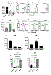
Fig. 5.. Effect of Ent on mitochondrial ROS generation.
ROS generation was measured in mitochondria isolated from the livers of BL6 mice using the redox-sensitive fluorescent probe 2’,7’-dichlorofluorescein-diacetate (DCFH-DA). Isolated mitochondria were incubated with DCFH-DA (5 μM) at 37°C for 10 min. The stained mitochondria (0.2 mg protein) were incubated with either Ent (10 μM) or FeCl3 (10 μM) or Ent+FeCl3 (1:1 ratio) and ROS generation was measured using fluorescence microplate reader for 30 min (excitation 488 nm and emission 525 nm). (A) Bar graphs showing the dose dependent effects of Ent on mitochondrial ROS generation. (B) The line graphs were represented as the DCF fluorescence over time. (C) Bar graphs represent the DCF formed/min/mg protein at the end point. (D) The DCFH stained mitochondria (0.2 mg protein) were incubated with either Ent (1–10 μM), FeCl3 (1–10 μM) or Ent+FeCl3 (1:1 ratio) and ROS generation was measured. (E) The mitochondria were incubated with either Ent, DFO, ferrichrome or pyoverdine (0–10 μM) and ROS generation was measured as DCF formed/min/mg protein. In vitro assays were performed in triplicates and are representative of three independent experiments. Results are expressed as mean ± SEM. * p< 0.05.

References

    1. Lui GY, Kovacevic Z, Richardson V, Merlot AM, Kalinowski DS, Richardson DR, Targeting cancer by binding iron: Dissecting cellular signaling pathways, Oncotarget 6(22) (2015) 18748–79. - PMC - PubMed
    1. Baudino TA, Targeted Cancer Therapy: The Next Generation of Cancer Treatment, Curr Drug Discov Technol 12(1) (2015) 3–20. - PubMed
    1. Urruticoechea A, Alemany R, Balart J, Villanueva A, Vinals F, Capella G, Recent advances in cancer therapy: an overview, Curr Pharm Des 16(1) (2010) 3–10. - PubMed
    1. Leoh LS, Kim YK, Candelaria PV, Martinez-Maza O, Daniels-Wells TR, Penichet ML, Efficacy and Mechanism of Antitumor Activity of an Antibody Targeting Transferrin Receptor 1 in Mouse Models of Human Multiple Myeloma, J Immunol 200(10) (2018) 3485–3494. - PMC - PubMed
    1. Richardson DR, Ponka P, The molecular mechanisms of the metabolism and transport of iron in normal and neoplastic cells, Biochim Biophys Acta 1331(1) (1997) 1–40. - PubMed

Publication types