

Developmental trajectories of the human embryologic brain regions
- PMID: 31228595
- PMCID: PMC6697108
- DOI: 10.1016/j.neulet.2019.134342
Developmental trajectories of the human embryologic brain regions
Abstract
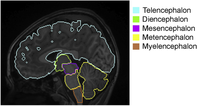
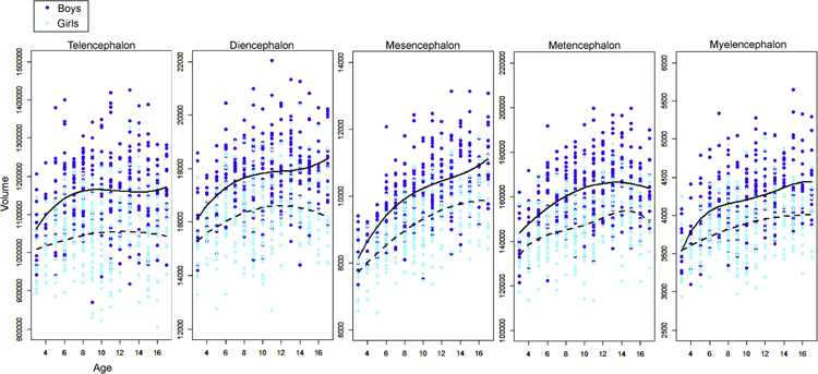
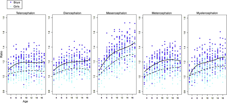
Vertebrate brains commonly consist of five basic embryologic anatomical regions: telencephalon; diencephalon; mesencephalon; metencephalon; and myelencephalon. The proportions of these regions vary widely across species and developmental stages. Investigation of their growth trajectories, therefore, has the potential to provide an understanding of the substrates of inter-species variation in neuroanatomy and function. To investigate the volumetric growth trajectories, structural magnetic resonance imaging (MRI) scans obtained from 618 healthy children (334 boys, 284 girls; ages 3-17 years old) were parcellated into five regions for the volume quantification. The sex- and region-specific growth trajectories were identified, and the most active growth was seen in the mesencephalon for both boys and girls. Whether similar regional growth patterns are seen in other species, or whether such patterns are related to evolution, are important questions that must be elucidated in the future.
Keywords: Atlas; Child; Diencephalon; Magnetic resonance imaging; Mesencephalon; Metencephalon; Myelencephalon; Telencephalon.
Copyright © 2019 Elsevier B.V. All rights reserved.
Figures
References
-
- Akaike H, [Data analysis by statistical models], No To Hattatsu 24 (1992) 127–133. - PubMed
-
- Andersen SL, Trajectories of brain development: point of vulnerability or window of opportunity?, Neurosci Biobehav Rev 27 (2003) 3–18. - PubMed
-
- Angold A, Costello EJ, Worthman CM, Puberty and depression: the roles of age, pubertal status and pubertal timing, Psychol Med 28 (1998) 51–61. - PubMed
-
- Bayer SA, Altman J, The human brain during the early first trimester, CRC ;Taylor & Francis distributor, Boca Raton, Fla. London, 2008, 522 p. pp.
-
- Brodal A, Neurological anatomy in relation to clinical medicine, Oxford University Press, New York, 1981, xvii, 1053 p. pp.
Publication types
MeSH terms
Grants and funding
LinkOut - more resources
Full Text Sources
