. 2019;8(3):513-526.
doi: 10.1016/j.jcmgh.2019.06.004. Epub 2019 Jun 19.

Generation of Human iPSC-Derived Intestinal Epithelial Cell Monolayers by CDX2 Transduction

Affiliations

Generation of Human iPSC-Derived Intestinal Epithelial Cell Monolayers by CDX2 Transduction

Kazuo Takayama et al. Cell Mol Gastroenterol Hepatol. 2019.

Abstract

Background & aims: To develop an effective and safe orally administered drug, it is important to predict its intestinal absorption rate, intestinal first-pass effect, and drug-drug interactions of orally administered drugs. However, there is no existing model to comprehensively predict the intestinal pharmacokinetics and drug-response of orally administered drugs. In this study, we attempted to generate homogenous and functional intestinal epithelial cells from human induced pluripotent stem (iPS) cells for pharmaceutical research.

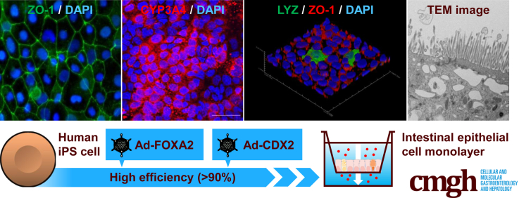
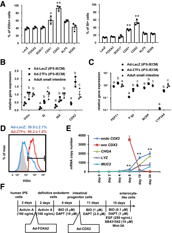
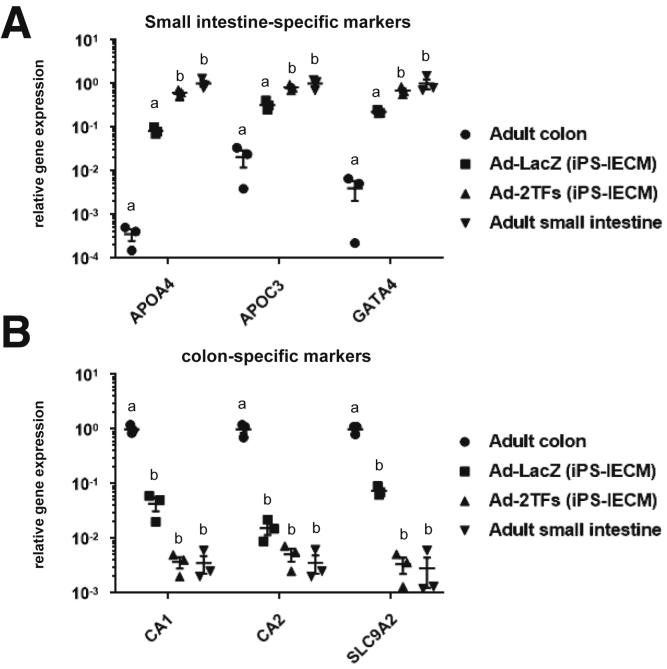
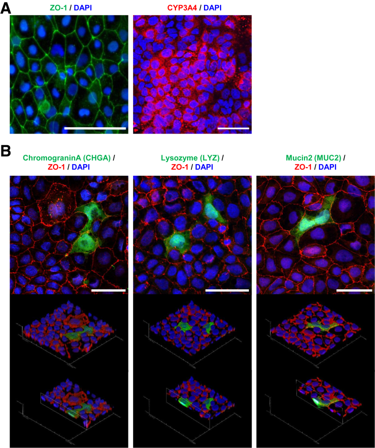
Methods: We generated almost-homogenous Villin- and zonula occludens-1 (ZO1)-positive intestinal epithelial cells by caudal-related homeobox transcription factor 2 (CDX2) transduction into human iPS cell-derived intestinal progenitor cells.

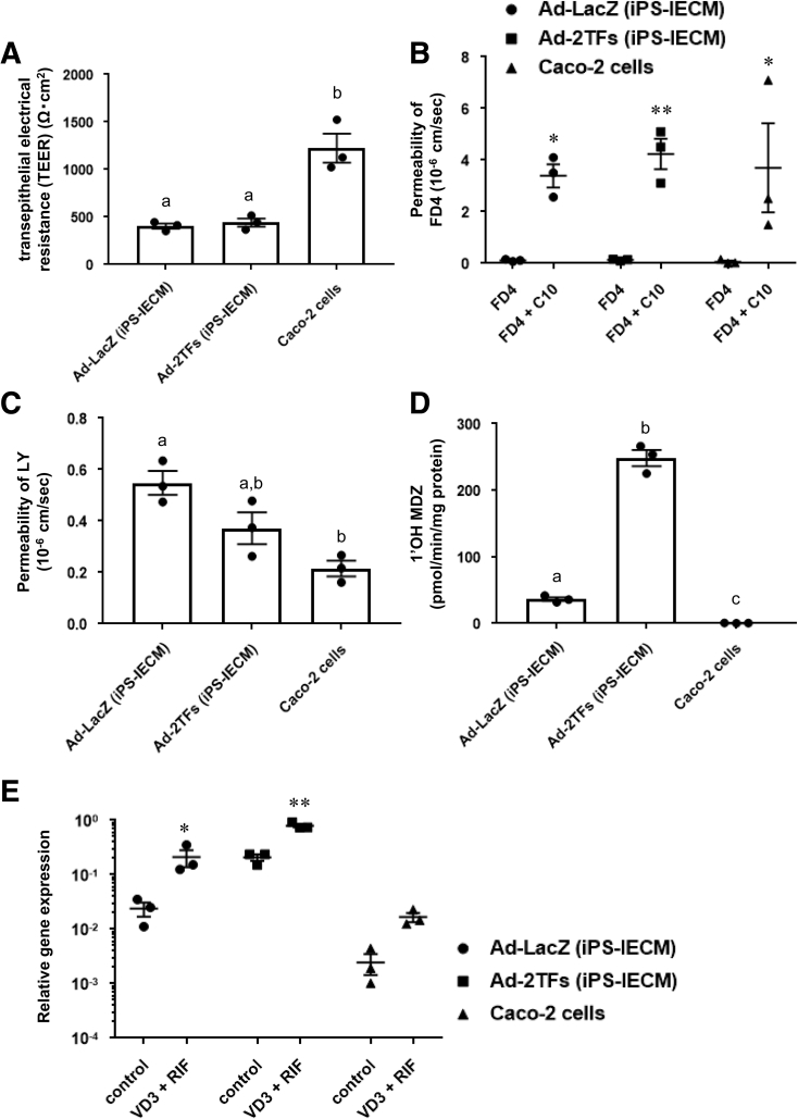
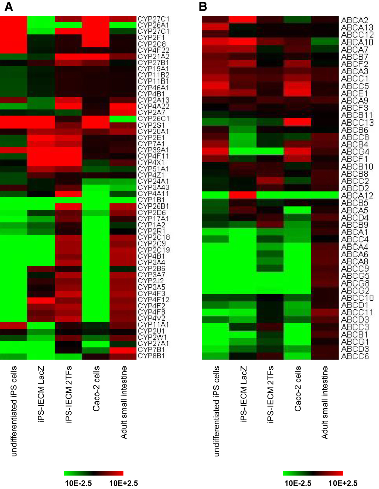
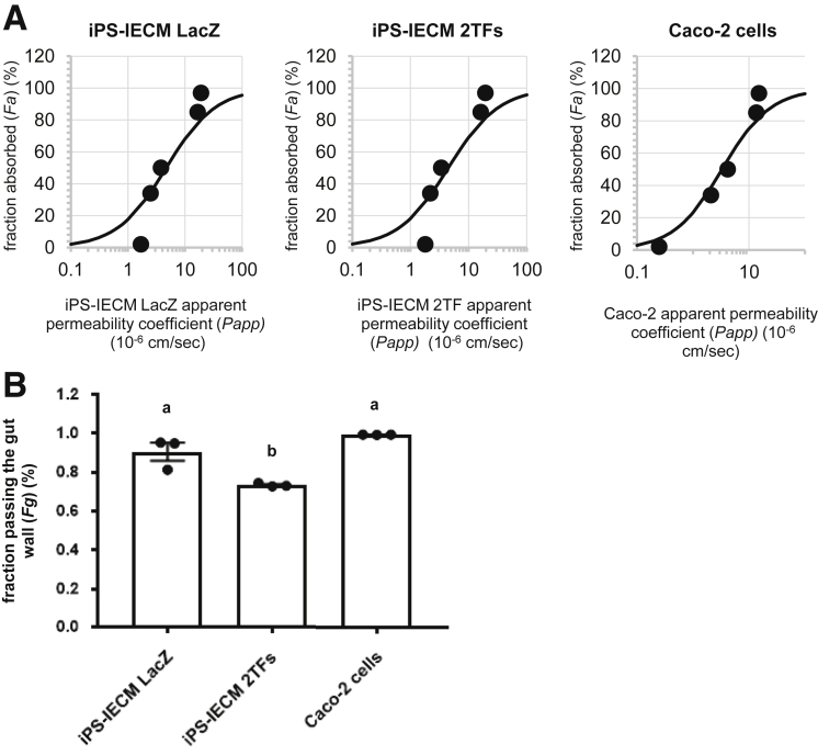
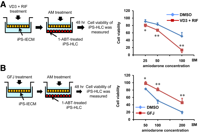
Results: The drug absorption rates in human iPS cell-derived intestinal epithelial cell monolayers (iPS-IECM) were highly correlated with those in humans (R2=0.91). The expression levels of cytochrome P450 (CYP) 3A4, a dominant drug-metabolizing enzyme in the small intestine, in human iPS-IECM were similar to those in human small intestine in vivo. In addition, intestinal availability in human iPS-IECM (the fraction passing the gut wall: Fg=0.73) was more similar to that in the human small intestine in vivo (Fg=0.57) than to that in Caco-2 cells (Fg=0.99), a human colorectal adenocarcinoma cell line. Moreover, the drug-drug interaction and drug-food interaction could be observed by using our human iPS-IECM in the presence of an inducer and inhibitor of CYP3A4, i.e., rifampicin and grape fruit juice, respectively.

Conclusion: Taking these results together, we succeeded in generating the human iPS-IECM that can be applied to various intestinal pharmacokinetics and drug-response tests of orally administered drugs.

Keywords: Adenovirus; CYP3A4; Differentiation; Intestinal First-Pass Effect.


Figures

None
Graphical abstract
Figure 1
Figure 1
Intestinal differentiation was promoted by CDX2 transduction. (A) Human iPSCs were differentiated into the intestinal progenitor cells as described in the Materials and Methods section. At day 8 of the differentiation, the intestinal progenitor cells were transduced with 3000 VPs/cell of Ad-TFs for 1.5 hours. Ad-TF–transduced intestinal progenitor cells were differentiated into intestinal epithelial cells as described in the Materials and Methods section. At day 34 of the differentiation, the percentage of Villin+ (left) and SI+ (right) cells was examined by fluorescence-activated cell sorting (FACS) analysis. Statistical significance was evaluated by 1-way analysis of variance followed by Bonferroni’s post hoc tests (*P < .05, **P < .01, vs Ad-LacZ–transduced cells). (B) The gene expression levels of Villin, SI, ISX, and CDX2 in Ad-LacZ–transduced cells, Ad-FOXA2-CDX2 (2TFs)–transduced cells, fetal small intestine, and adult small intestine were examined by real-time RT-PCR. The gene expression levels in the small intestine were taken as 1.0. (C) The gene expression levels of peptide transporter 1, P-glycoprotein, breast cancer resistance protein, and CYP3A4 in Ad-LacZ–transduced cells, Ad-FOXA2-CDX2 (2TFs)–transduced cells, fetal small intestine, and adult small intestine were examined by real-time RT-PCR. The gene expression levels in the small intestine were taken as 1.0. (B, C) Statistical significance was evaluated by 2-way analysis of variance followed by Tukey’s post hoc tests. Groups that do not share the same letter are significantly different from each other (P < .05). (D) The percentage of Villin+ cells in Ad-LacZ–transduced cells and Ad-2TF–transduced cells was examined by FACS analysis. (E) The endogenous and exogenous CDX2, CHGA, LYZ, and MUC2 expression levels were measured by absolute real-time RT-PCR. Statistical significance was evaluated by Student’s t test (endogenous CDX2 vs exogenous CDX2; **P < .01). (F) The procedure for intestinal differentiation is presented schematically. Details of the intestinal differentiation procedure are described in the Materials and Methods section. All data are shown as the mean ± SE of 3 independent differentiation experiments.
Figure 2
Figure 2
The gene expression analysis of small intestine- and colon-specific markers. Human iPSCs were differentiated into the intestinal epithelial cell monolayers as described in the Materials and Methods section. The gene expression levels of (A) small intestine-specific markers and (B) colon-specific markers in Ad-LacZ–transduced cells, Ad-FOXA2-CDX2 (2TFs)–transduced cells, adult colon, and adult small intestine were examined by real-time RT-PCR. The gene expression levels in (A) the adult small intestine and (B) adult colon were taken as 1.0. Statistical significance was evaluated by 2-way analysis of variance followed by Tukey’s post hoc tests. Groups that do not share the same letter are significantly different from each other (P < .05). All data are shown as the mean ± SE of 3 independent differentiation experiments.
Figure 3
Figure 3
Immunostaining analysis of human iPS-IECMs. Human iPSCs were differentiated into the intestinal epithelial cell monolayers on the chamber as described in the Materials and Methods section. (A) The ZO-1 and CYP3A4 expression levels were examined by immunostaining analysis. Nuclei were counterstained with DAPI. Scale bars = 50 μm. (B) Immunostaining analysis of CHGA, LYZ, MUC2, and ZO-1 was performed in the human iPS-IECMs. Nuclei were stained with DAPI (blue). Scale bars = 50 μm.
Figure 4
Figure 4
Barrier and metabolic functions of human iPSC–derived intestinal epithelial cell monolayers. (A) TEER values of the Ad-LacZ–transduced iPS-IECMs, Ad-2TF–transduced iPS-IECMs, and Caco-2 monolayers were measured by Millicell-ERS2. (B) Apical-to-basolateral permeability of FD4 across the Ad-LacZ–transduced monolayers, Ad-2TF–transduced monolayers, and Caco-2 monolayers was measured in the presence or absence of C10 (an absorption-enhancing agent). (C) Apical-to-basolateral permeability of LY across the Ad-LacZ–transduced iPS-IECMs, Ad-2TF–transduced iPS-IECMs, and Caco-2 monolayers was measured. (D) The CYP3A4-mediated drug metabolizing capacities in the Ad-LacZ–transduced iPS-IECMs, Ad-2TF–transduced iPS-IECMs, and Caco-2 monolayers were evaluated by quantifying the metabolites of the CYP3A4 substrate, MDZ. The quantity of metabolites, 1′-OH MDZ, was measured by UPLC-MS/MS. (E) To examine CYP3A4 induction potency, the cells were treated with 100-nM VD3 for 21 days and 20-μM RIF for 2 days. The gene expression levels of CYP3A4 were measured by real-time RT-PCR. Controls were treated with DMSO (final concentration 0.1%). The gene expression levels in the human small intestine were taken as 1.0. (A, C, D) Statistical significance was evaluated by 1-way analysis of variance followed by Tukey’s post hoc tests. Groups that do not share the same letter are significantly different from each other (P < .05). (B, E) Statistical significance was evaluated by 2-way analysis of variance followed by Bonferroni’s post hoc tests (*P < .05, **P < .01, FD4 vs FD4 + C10 and control vs VD3 + RIF, respectively). The results are shown as the mean ± SE of 3 independent differentiation experiments.
Figure 5
Figure 5
The gene expression profile of CYPs and ABC transporters in human iPS-IECMs. Human iPSCs were differentiated into the intestinal epithelial cell monolayers as described in the Materials and Methods section. The global gene expression analysis was performed in undifferentiated human iPSCs, human Ad-LacZ–transduced iPS-IECMs, human Ad-2TF–transduced iPS-IECMs, Caco-2 cells, and the human adult small intestine in vivo. Heatmap analyses of (A) CYPs and (B) ABC transporters are shown.
Figure 6
Figure 6
Human iPSC–derived intestinal epithelial cell monolayers can be utilized in evaluating Fg value. Human iPSCs were differentiated into the intestinal epithelial cell monolayers on the chamber as described in the Materials and Methods section. (A) The apical-to-basolateral permeability of antipyrine, metoprolol, fexofenadine, atenolol, and pravastatin across the Ad-2TF–transduced iPS-IECMs was measured. The data on the human proximal jejunum permeability of antipyrine, metoprolol, fexofenadine, atenolol, and pravastatin in vivo are cited from previously published papers., , (B) To examine the in vitro Fg in the Ad-LacZ– and Ad-2TF–transduced iPS-IECMs and Caco-2 cells, the CYP3A4-mediated drug metabolizing capacities were evaluated by quantifying the MDZ and 1′-OH MDZ in the apical chamber, basolateral chamber, and cells. The Fg was calculated as described in the Materials and Methods section. Statistical significance was evaluated by 1-way analysis of variance followed by Tukey’s post hoc tests. Groups that do not share the same letter are significantly different from each other (P < .05). The results are shown as the mean ± SE of 3 independent differentiation experiments.
Figure 7
Figure 7
Human iPSC–derived intestinal epithelial cell monolayers can be utilized in predicting drug-drug interactions. Human iPSCs were differentiated into the intestinal epithelial cell monolayers on the chamber as described in the Materials and Methods section. (A) To induce CYP3A4 activity, Ad-2TF–transduced iPS-IECMs were cultured with VD3 for 21 days and RIF for 2 days. Ad-2TF–transduced iPS-IECMs and 1-aminobenzotriazole–treated iPSC-derived hepatocyte-like cells were co-cultured with amiodarone-containing medium for 48 hours. The cell viability of the hepatocyte-like cells was measured. The cell viability of nontreated cells was taken as 100. Statistical significance was evaluated by repeated 2-way analysis of variance followed by Bonferroni’s post hoc tests (*P < .05, **P < .01, DMSO vs VD3 + RIF). (B) To inhibit CYP3A4 activity, Ad-2TF–transduced iPS-IECMs were cultured with 6′,7′-dihydroxybergamottin and bergamottin (GFJ) for 48 hours. Ad-2TF–transduced iPS-IECMs and hepatocyte-like cells were co-cultured with Amiodarone-containing medium for 48 hour. The cell viability of the hepatocyte-like cells was measured. The cell viability of nontreated cells was taken as 100. Statistical significance was evaluated by repeated 2-way analysis of variance followed by Bonferroni’s post hoc tests (*P < .05, **P < .01, DMSO vs GFJ). The results are shown as the mean ± SE of 3 independent differentiation experiments.

References

    1. Watkins P.B., Wrighton S., Schuetz E., Molowa D.T., Guzelian P. Identification of glucocorticoid-inducible cytochromes P-450 in the intestinal mucosa of rats and man. J Clin Invest. 1987;80:1029–1036. - PMC - PubMed
    1. Paine M.F., Khalighi M., Fisher J.M., Shen D.D., Kunze K.L., Marsh C.L., Perkins J.D., Thummel K.E. Characterization of interintestinal and intraintestinal variations in human CYP3A-dependent metabolism. J Pharmacol Exp Ther. 1997;283:1552–1562. - PubMed
    1. Thummel K.E., Kunze K.L., Shen D.D. Enzyme-catalyzed processes of first-pass hepatic and intestinal drug extraction. Adv Drug Deliv Rev. 1997;27:99–127. - PubMed
    1. Fromm M.F., Busse D., Kroemer H.K., Eichelbaum M. Differential induction of prehepatic and hepatic metabolism of verapamil by rifampin. Hepatology. 1996;24:796–801. - PubMed
    1. Gomez D.Y., Wacher V.J., Tomlanovich S.J., Hebert M.F., Benet L.Z. The effects of ketoconazole on the intestinal metabolism and bioavailability of cyclosporine. Clin Pharm Ther. 1995;58:15–19. - PubMed

Publication types

MeSH terms