. 2019 Oct:81:317-328.
doi: 10.1016/j.bbi.2019.06.028. Epub 2019 Jun 20.

Neurotoxicity to dopamine neurons after the serial exposure to alcohol and methamphetamine: Protection by COX-2 antagonism

Affiliations

Neurotoxicity to dopamine neurons after the serial exposure to alcohol and methamphetamine: Protection by COX-2 antagonism

Amanda L Blaker et al. Brain Behav Immun. 2019 Oct.

Abstract

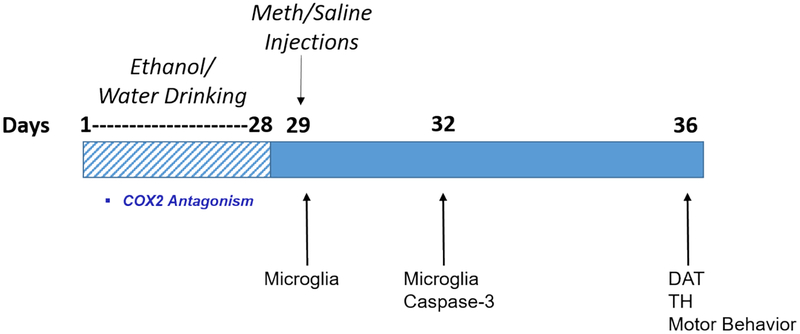
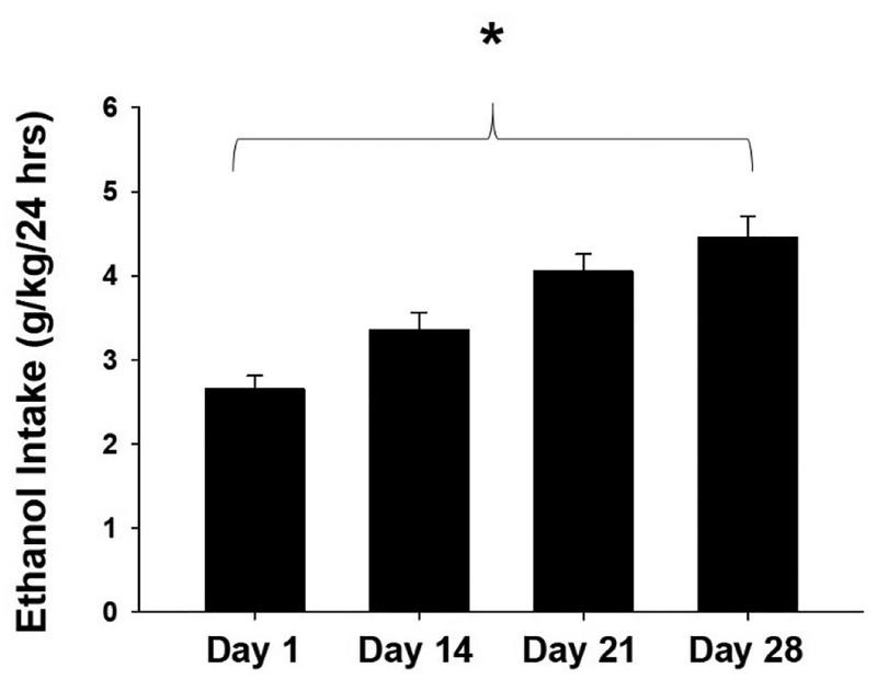
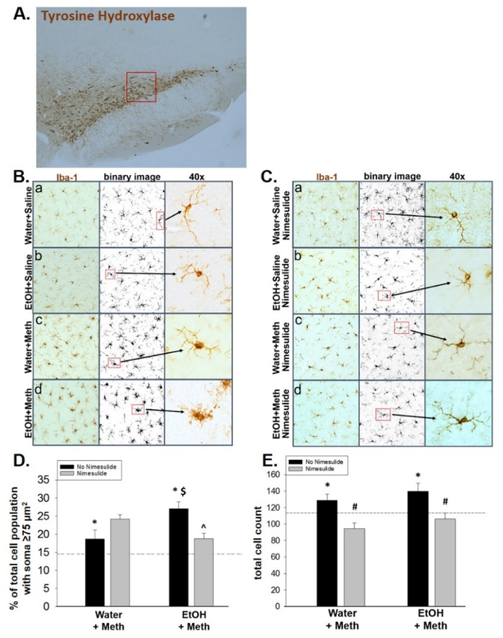
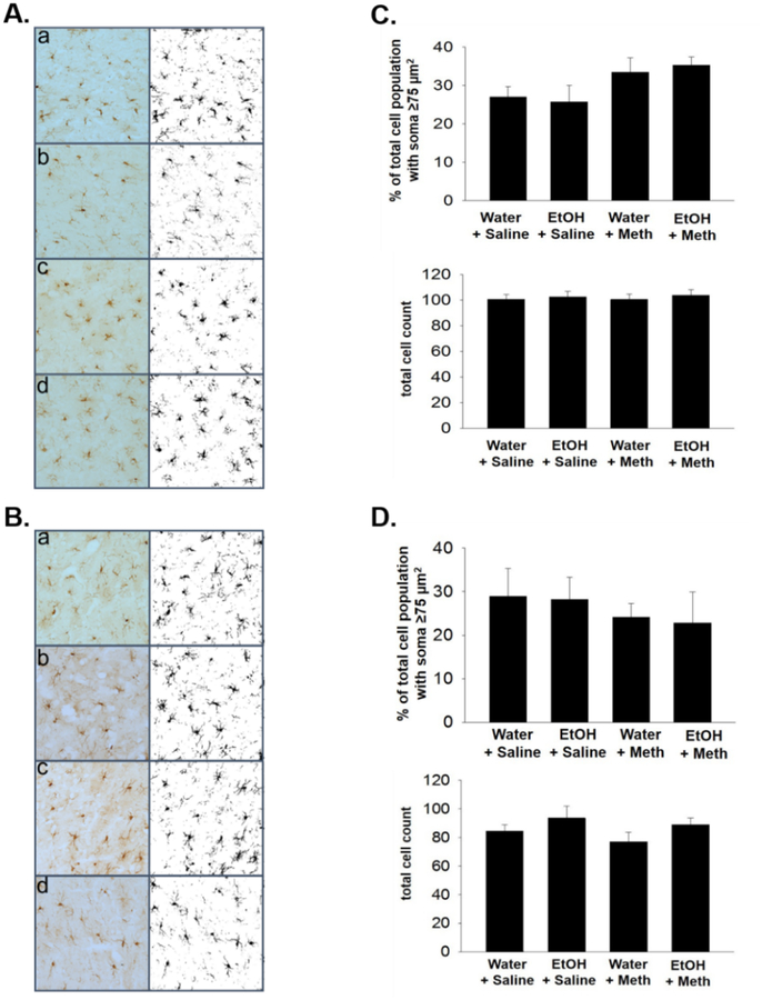
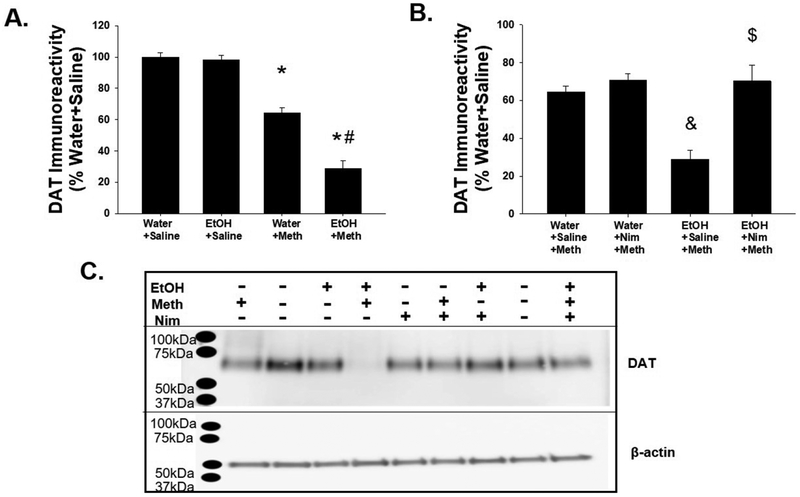
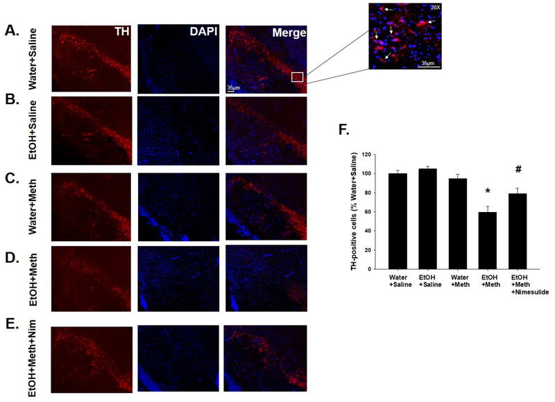
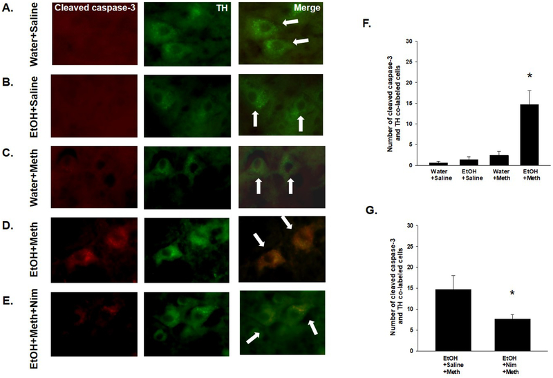
A significant co-morbidity exists between alcohol and methamphetamine (Meth) in humans but the consequences and mechanisms underlying their co-morbid effects remain to be identified. A consequence associated with the abuse of either alcohol or Meth involves inflammation but little is known about the role of inflammation in a possible neurotoxicity arising from their co-exposure. Sprague Dawley rats were allowed 28 days of intermittent, voluntary access to 10% ethanol (EtOH) followed by a neurotoxic binge administration of Meth. EtOH drinking followed by Meth increased microglial cell counts and produced morphological changes in microglia of the substantia nigra pars compacta 2 h after Meth administration that were distinct from those produced by either EtOH or Meth alone. These effects preceded the activation of cleaved caspase-3 in dopamine cell bodies, as well as decreases in tyrosine hydroxylase (TH) immunoreactivity in the substantia nigra and dopamine transporter (DAT) immunoreactivity in the striatum measured at 7 days after Meth. Intervention with a selective COX-2 inhibitor during EtOH drinking prevented the changes in microglia, and attenuated the increase in cleaved caspase-3, and decreases in TH and DAT after Meth administration. Furthermore, motor dysfunction measured by a rotarod test was evident but only in rats that were exposed to both EtOH and Meth. The motor dysfunction was ameliorated by prior inhibition of COX-2 during EtOH drinking. The exaggerated neurochemical and behavioral deficits indicate that the comorbidity of EtOH and Meth induces a degeneration of the nigrostriatal pathway and support the role of inflammation produced by EtOH drinking that primes and mediates the neurotoxic consequences associated with the common co-morbidity of these drugs.


Figures

Fig. 1.
Fig. 1.
A timeline describing the experimental paradigm and experimental measures.
Fig. 2.
Fig. 2.
Drinking behavior in Sprague Dawley rats over 28 days. Rats increased EtOH intake (g/kg/24 hrs) over time (*p < 0.001; n = 97). Data are presented as mean ± SEM.
Fig. 3.
Fig. 3.
EtOH+Meth changes morphology and number of Iba-1 immunoreactive cells 2hr after Meth and this is mediated by COX-2. A) The region of interest within the SNpc is outlined in red (5X). Representative images of Iba-1 immunoreactivity in rats with B) no nimesulide and C) nimesulide intervention during EtOH drinking. Images are at 20x and 40x. D) Meth-treated rats displayed significantly larger % of total cells with large cell bodies compared to saline-treated rats (n = 6–12/group, * p < 0.05). This was further enhanced by EtOH (n = 3–12/group, $p < 0.05) and was blocked by nimesulide (^p < 0.05). E) Meth treatment significantly increased total number of microglia (n = 6–12/group, *p < 0.05) and this was also blocked by nimesulide (n = 3–12/group, #p < 0.05). Results are presented as mean ± SEM. The dotted line represents the mean of the Water+No Nimesulide+Saline (D–E).
Fig. 4.
Fig. 4.
Iba-1 immunoreactive cells of the SNpc lose morphological complexity 72 hr post Meth binge. A) Representative images of Iba-1 stained sections from animals at 72 hr post Meth binge from a) Water+Saline, b) EtOH+Saline, c) Water+Meth, and d) EtOH+Meth treatment groups. Images are at 20x. B–C) Analysis of Iba-1 stained cells within the SNpc at 72 hr post Meth binge indicates that changes in B) cell soma size measurements, and C) the total number of cells identified at 2 h, are no longer present. n = 3–6/group
Fig. 5.
Fig. 5.
Iba-1 immunoreactive cells of the striatum show no changes in morphology after treatment. Representative images of Iba-1 stained striatal sections from animals at A) 2h and B) 72h post Meth binge from a) Water+Saline, b) EtOH+Saline, c) Water+Meth, and d) EtOH+Meth treatment groups are shown. An analysis of Iba-1 stained cells in the striatum at C) 2h and D) 72h post Meth binge indicates no effect of EtOH, Meth, or the combination, at these time points. Results are presented as mean ± SEM. n = 3–12/group
Fig. 6.
Fig. 6.
COX-2 inhibition during EtOH drinking prevents enhanced Meth-induced DAT depletions. A) DAT was significantly decreased in Meth-treated vs. saline-treated rats (n = 6–8/group; *p < 0.05) and further decreased in rats previously exposed to EtOH (#p < 0.05 vs. Water+Meth). B) Nimesulide treatment during EtOH drinking blocked the synergistic decreases in DAT after EtOH+Meth (n = 6–8/group; &p < 0.05 vs. Water+Saline+Meth; $p < 0.05 vs. EtOH+Saline+Meth). C) Representative image of Western blot for DAT with β-actin as the loading control.
Fig. 7.
Fig. 7.
TH+ immunoreactivity is decreased in the SNpc 7 days after EtOH+Meth. TH+ immunoreactivity in the SNpc is shown in A) Water+Saline, B) EtOH+Saline, C) Water+Meth, D) EtOH+Meth, and E) EtOH+Meth+Nim treatment groups. F) EtOH+Meth rats displayed significantly less TH+ cells than all other groups (n = 6–8/group; *p < 0.05). Meth rats treated with nimesulide during EtOH drinking did not show the same extent of decreased TH-positive cells in the SNpc compared to EtOH+Meth (#p < 0.05). Data are expressed as mean ± SEM.
Fig. 8.
Fig. 8.
EtOH+Meth induces apoptosis in dopaminergic neurons in the SNpc. Representative images for cleaved caspase-3 and TH immunofluorescence are provided for A) Water+Saline, B) EtOH+Saline, C) Water+Meth, and D) EtOH+Meth. F) EtOH+Meth significantly increased the number of cleaved caspase-3 and TH co-labeled cells in the SNpc (*p < 0.05 vs. all groups). E) A subset of rats was treated with nimesulide during EtOH drinking and this attenuated the number of apoptotic cells in the SNpc (G). *p < 0.05. n = 5–8/group. Data are represented as mean ± SEM.
Fig. 9.
Fig. 9.
EtOH+Meth decrease motor function at 7 days after Meth. A) 2-way ANOVA showed a significant interaction between EtOH and Meth (n = 8–14/group; p < 0.001). Tukey post-hoc analyses showed that EtOH+Meth rats fell off the rotarod sooner than their EtOH-alone or Meth-alone counterparts (*p < 0.001). B) Rats treated with nimesulide in addition to EtOH+Meth did not exhibit motor deficits compared to the EtOH+Meth with saline vehicle (Student’s t-test; n = 8–9/group; *p < 0.05).

Similar articles

Cited by

References

    1. Anneken JH, et al., 2013. MDMA increases glutamate release and reduces parvalbumin-positive GABAergic cells in the dorsal hippocampus of the rat: role of cyclooxygenase. J Neuroimmune Pharmacol. 8, 58–65. - PMC - PubMed
    1. Ares-Santos S, et al., 2014. Methamphetamine causes degeneration of dopamine cell bodies and terminals of the nigrostriatal pathway evidenced by silver staining. Neuropsychopharmacology. 39, 1066–80. - PMC - PubMed
    1. Banks WA, 2005. Blood-brain barrier transport of cytokines: a mechanism for neuropathology. Curr Pharm Des. 11, 973–84. - PubMed
    1. Barcia C, et al., 2012. ROCK/Cdc42-mediated microglial motility and gliapse formation lead to phagocytosis of degenerating dopaminergic neurons in vivo. Sci Rep. 2, 809. - PMC - PubMed
    1. Barcia C, et al., 2004. Evidence of active microglia in substantia nigra pars compacta of parkinsonian monkeys 1 year after MPTP exposure. Glia. 46, 402–9. - PubMed

Publication types

MeSH terms