Review
. 2019 Jun:58:142-152.
doi: 10.1016/j.ceb.2019.05.001. Epub 2019 Jun 20.

Two major mechanisms of chromosome organization

Affiliations
Review

Two major mechanisms of chromosome organization

Leonid A Mirny et al. Curr Opin Cell Biol. 2019 Jun.

Abstract

The spatial organization of chromosomes has long been connected to their polymeric nature and is believed to be important for their biological functions, including the control of interactions between genomic elements, the maintenance of genetic information, and the compaction and safe transfer of chromosomes to cellular progeny. chromosome conformation capture techniques, particularly Hi-C, have provided a comprehensive picture of spatial chromosome organization and revealed new features and elements of chromosome folding. Furthermore, recent advances in microscopy have made it possible to obtain distance maps for extensive regions of chromosomes (Bintu et al., 2018; Nir et al., 2018 [2••,3]), providing information complementary to, and in excellent agreement with, Hi-C maps. Not only has the resolution of both techniques advanced significantly, but new perturbation data generated in the last two years have led to the identification of molecular mechanisms behind large-scale genome organization. Two major mechanisms that have been proposed to govern chromosome organization are (i) the active (ATP-dependent) process of loop extrusion by Structural Maintenance of Chromosomes (SMC) complexes, and (ii) the spatial compartmentalization of the genome, which is likely mediated by affinity interactions between heterochromatic regions (Falk et al., 2019 [76••]) rather than by ATP-dependent processes. Here, we review existing evidence that these two processes operate together to fold chromosomes in interphase and that loop extrusion alone drives mitotic compaction. We discuss possible implications of these mechanisms for chromosome function.


Conflict of interest statement

Conflict of Interest

The authors declare no conflict of interest.

Figures
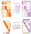
Figure 1.
Figure 1.. Hallmark patterns in Hi-C maps: compartmentalization and TADs
(a) Compartmentalization. Pattern in Hi-C (left): The characteristic checkerboard pattern of alternating zones of contact enrichment and depletion spanning the intra- and interchromosomal areas of the genome-wide Hi-C map. Chromosomes 16 and 17 from HFFc6 human fibroblast cells are shown (data from https://data.4dnucleome.org/). Interpretation (middle): Genomic intervals of different type exhibit differential affinities that cause them to segregate spatially in the nucleus. At the coarsest level, this can be broken down into two compartmental types: A (active, magenta) and B (inactive, blue) chromatin. Schematic (right): A simplified annotation, where colored rectangles represent zones of homotypic contact enrichment and blank rectangles represent zones of heterotypic contact depletion. The colors in the margin represent the A/B identity along the chromosomes. The translucence of trans and inter-arm areas of the map represents the different baseline contact frequencies associated with territoriality of chromosomes and chromosome arms as seen in Hi-C on the left. (b) TADs and associated cohesin-dependent patterns. Pattern in Hi-C (left): Areas along the diagonal of the contact map at shorter genomic ranges (normally <3Mb) show elevated contact frequencies and exhibit characteristic features such as stripes, peaks and peak grids based typically at CTCF sites. In this example region, the lower triangle of the heatmap uses a linear color scale that highlights the details of TAD patterns, while the upper triangle of the heatmap uses a logarithmic color scale that enables one to observe TAD patterns superimposed upon compartmental domains and their associated checkered zones. Interpretation (middle): An ATP-dependent process of loop extrusion by cohesin complexes locally compacts chromosomes and acts independently of the forces driving compartmentalization. Genomically localized barrier elements constrain the range by which loop extrusion can mediate contacts, which gives rise to TAD-associated patterns of contact enrichment [12]. Schematic (right): A simplified depiction of the range of loop extrusion-mediated contact enrichment (dark grey squares) limited by barriers depicted in red on the diagonal and within the yellow bars in the margin. In the background, compartmental domains and zones of enrichment and depletion are colored as in (a). The lower triangle is translucent to reflect the dynamic range of signal when using a linear color scale as in the Hi-C example on the left.
Figure 2.
Figure 2.. TAD formation by loop extrusion.
Bottom: diagrams of population contact frequency maps of a genomic region. (a) Basal level of contact frequency in the absence of loop extrusion. (b) Loop extrusion creates additional short-range contacts, further compacting chromatin. No extrusion-dependent domains are visible in Hi-C in the absence of barriers. (c) Extrusion barriers limit the additional extrusion-mediated contacts to intervals delimited by barriers, thus giving rise to TADs, while the contact frequency between TADs is lower. Loop extrusion forms TADs by mediating additional intra-TAD contacts, rather than by insulating spontaneous spatial contacts. Stripes and peaks also naturally emerge when cohesins are stopped by CTCF at TAD borders. Top: cartoon depicting a single chromatin fiber (a) without loop extrusion; (b) subject to loop extrusion; (c) subject to loop extrusion with barriers. Symbols are drawn both on the cartoons to illustrate the extrusion machinery on chromatin and on top of the map to indicate the possible instantaneous positions of loop extruders in the example conformation above. Yellow circles depict loop extruders, and red octagons depict extrusion barriers.
Figure 3.
Figure 3.. Two major mechanisms of chromosome organization.
Nucleosomal arrays create a chromatin fiber that is folded differently in interphase and metaphase. In mitosis, loop extrusion by condensins leads to the formation of a dense array of loops: condensins form a scaffold with loop emanating from it. In interphase, loop extrusion is performed by cohesin forming a sparse dynamic array of loops, with CTCFs forming barriers to extrusion. Heterochromatin and euchromatin are further compartmentalized in space by the attraction of heterochromatic regions to one another.

References

    1. Marko JF, Siggia ED: Stretching DNA. Macromolecules 1995, 28:8759–8770.
    1. Bintu B, Mateo LJ, Su J-H, Sinnott-Armstrong NA, Parker M, Kinrot S, Yamaya K, Boettiger AN, Zhuang X: Super-resolution chromatin tracing reveals domains and cooperative interactions in single cells. Science 2018, 362.

      (**) Distance maps obtained by microscopy show small distance for loci within, and larger between, TADs. Upon cohesin depletion, intra-TAD distances go up. This decompaction upon loss of cohesin is consistent with loop extrusion and was proposed in Fudenberg et al 2016.

    1. Nir G, Farabella I, Pérez Estrada C, Ebeling CG, Beliveau BJ, Sasaki HM, Lee SD, Nguyen SC, McCole RB, Chattoraj S, et al.: Walking along chromosomes with super-resolution imaging, contact maps, and integrative modeling. PLoS Genet 2018, 14:e1007872. - PMC - PubMed
    1. Korzybski A: Science and sanity: an introduction to non-Aristotelian systems and general semantics. The International Non-Aristotelian Library Pub. Co.; 1933.
    1. Rao SSP, Huntley MH, Durand NC, Stamenova EK, Bochkov ID, Robinson JT, Sanborn AL, Machol I, Omer AD, Lander ES, et al.: A 3D map of the human genome at kilobase resolution reveals principles of chromatin looping. Cell 2014, 159:1665–1680. - PMC - PubMed

Publication types

Substances