. 2019 Oct 1:461:21-30.
doi: 10.1016/j.canlet.2019.06.011. Epub 2019 Jun 20.

Frequent amplifications of ESR1, ERBB2 and MDM4 in primary invasive lobular breast carcinoma

Affiliations

Frequent amplifications of ESR1, ERBB2 and MDM4 in primary invasive lobular breast carcinoma

Lan Cao et al. Cancer Lett. .

Abstract

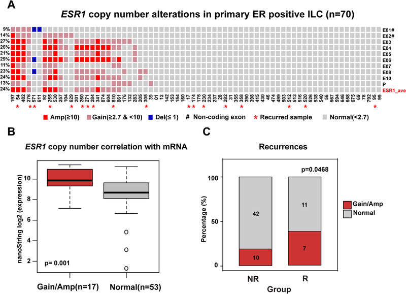
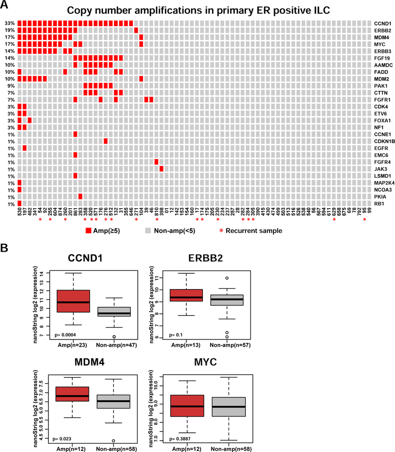
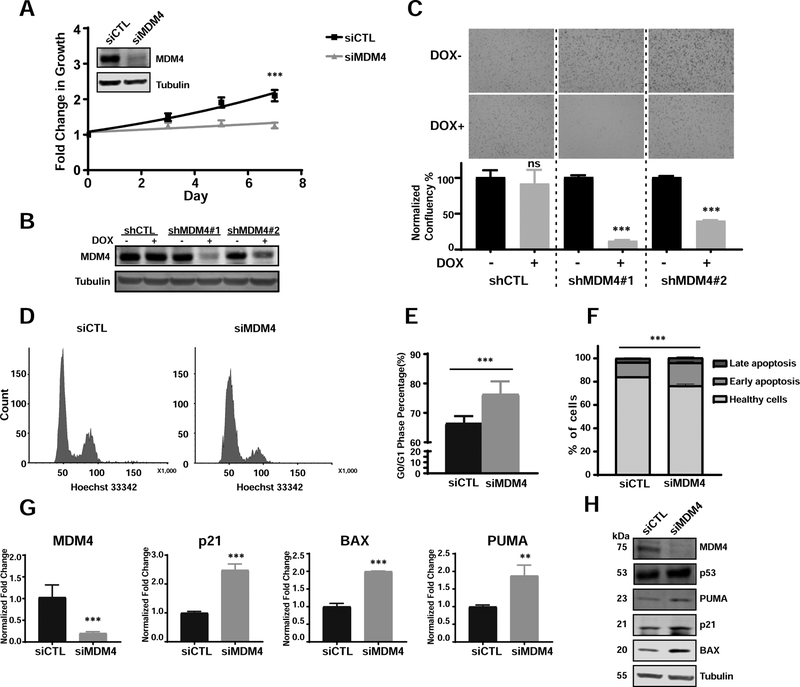
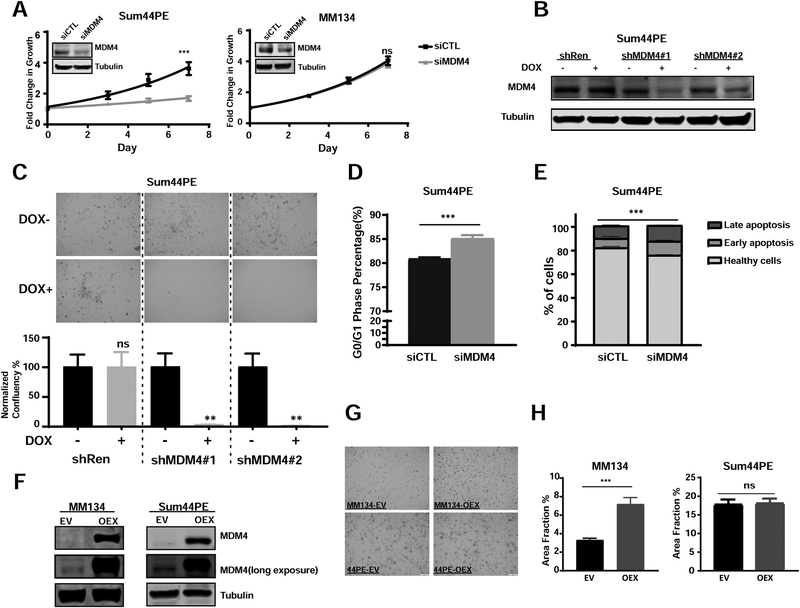
Invasive lobular carcinoma (ILC) is the second most common histological subtype of breast cancer following invasive ductal carcinoma (IDC). To identify potential genetic drivers of ILC progression, we used NanoString nCounter technology to investigate the DNA copy number (CN) in 70 well-curated primary ILC samples. We confirmed prior observations of frequent amplification of CCND1 (33%), and MYC (17%) in ILC, but additionally identified a substantial subset of ILCs with ESR1 and ERBB2 (19%) amplifications. Of interest, tumors with ESR1 CN gains (14%) and amplification (10%) were more likely to recur compared to those with normal CN. Finally, we observed that MDM4 (MDMX) was amplified in 17% of ILC samples. MDM4 knockdown in TP53 wild-type ILC cell lines caused increased apoptosis, decreased proliferation associated with cell cycle arrest, and concomitant activation of TP53 target genes. Similar effects were seen in TP53 mutant cells, indicting a TP53-independent role for MDM4 in ILC. To conclude, amplification of ESR1 and MDM4 are potential genetic drivers of ILC. These amplifications may represent actionable, targetable tumor dependencies, and thus have potential clinical implications and warrant further study.

Keywords: Amplification; ERBB2; ESR1; ILC; MDM4.


Conflict of interest statement

Conflict of interest: The authors declare they have no conflicts of interest with the contents of this article.

Figures

Figure 1.
Figure 1.. Frequent ESR1 copy number alterations in 70 primary ER positive ILC specimens.
A. Oncoprint visualization of ESR1 CN alterations in primary ER positive ILC. Levels of amplification and deletion are color-coded. Each column represents a single sample. Each row indicates the copy number call of the corresponding single exon probe. Frequencies of alterations are indicated to the left side of each row. Samples with subsequent recurrences are annotated with ‘*’ symbol. E01-E10: exons 1–10. Untranslated exons (E1 and E2) are annotated with ‘#’ symbol. P: Promoter probe. ESR1_ave: average copy number call of all probes. B. Box plot comparison of ESR1 mRNA expression measured by NanoString between normal and gain/amplified samples (Wilcoxon rank-sum test). C. Frequency of ESR1 CN amplifications for the non-recurrence (NR) and recurrence groups (R) (Chi-square test, one-side, p=0.0468). Numbers show the count of tumors for each group. Amp: amplification. Del: deletion.
Figure 2.
Figure 2.. CCND1, ERBB2, MDM4 and MYC are the most frequently amplified genes in ER positive primary ILC.
A. Oncoprint visualization of CN amplifications by genes (rows) and samples (columns). Frequencies of the amplifications are indicated to the left side of each row. Samples with later recurrence are annotated with ‘*’ symbol. B. Comparison of NanoString mRNA expression of most frequently amplified genes in the ILC cohort between amplified and non-amplified samples (Wilcoxon rank-sum test). Amp: amplification.
Figure 3.
Figure 3.. Targeting MDM4 in p53-functional breast cancer cell line BCK4 suppresses cell growth by inducing G0/G1 arrest and apoptosis.
A. Growth curves of BCK4 cells transiently transfected with siMDM4 vs siCTL (two-way ANOVA, ***p<0.001, n=2). MDM4 transient KD was confirmed by immunoblots shown top left. B. shRNA KD in BCK4 cells confirmed by immunoblot. DOX: doxycycline. C. Representative images from colony formation assays of BCK4 shCTL and shMDM4 cells. Quantifications of normalized confluency are shown below (data represent the average of biological triplicates ±SD; t-tests, ***p<0.001, n=2). D. Representative FACS figures of cell cycle analysis of siCTL and siMDM4 after knockdown for 4 days. E. Percentage of cells in G0/G1 phase in BCK4 siCTL vs siMDM4 after transfection for 4 days (t-tests, data represent the average of 8 biological replicates from three independent experiments ±SD). F. Apoptosis assay profiles in BCK4 cells transfected with siCTL or siMDM4 after 4 days (p value by t-test for total apoptotic cells in siCTL vs siMDM4, ***p<0.001, n=3). G. qPCR of p53 downstream targets in BCK4 cells after MDM4 downregulation (Data represent the average of biological triplicates ±SD; t-tests, **p<0.05, ***p<0.001). H. Immunoblots demonstrating induction of p53 target genes in BCK4 cells after MDM4 downregulation by siRNA.
Figure 4.
Figure 4.. MDM4 promotes cell cycle progression in mutant TP53 breast cancer cell lines.
A. Growth curves of Sum44PE (left) and MM134 (right) cells transiently transfected with siMDM4 vs siCTL (two-way ANOVA, ***p<0.001, ns: no significance, n=4). MDM4 knockdown was confirmed by immunoblots shown top left. B. shRNA KD in Sum44PE cells was confirmed by immunoblot. C. Representative images from colony formation assay in Sum44PE shCTL and shMDM4 cells. Quantification of normalized confluency are shown below (data represent the average of biological triplicates ±SD; t-tests, ***p<0.001, n=2). D. Percentage of cells in G0/G1 phase in Sum44PE siCTL vs siMDM4 after transfection for 4 days (t-tests, data represent the average of 6 biological replicates from two independent experiments ±SD). E. Apoptosis assay profiles in Sum44PE cells transfected with siCTL or siMDM4 after 4 days (p value by t-test for total apoptotic cells in siCTL vs siMDM4, ***p<0.001, n=2). F. Immunoblot validation of constitutive MDM4 overexpression in MM134 (left) and Sum44PE (right) cells. G. Representative images of MM134 (left) and Sum44PE (right) cells transfected with EV (empty vector) and OEX (MDM4 overexpresssion plasmid) after 18-days growth in a colony formation assay. H. Quantifications by area fraction of all the colonies in MM134 (left) and Sum44PE (right) (t-tests, ***p<0.001, n≥2)

References

    1. Martinez V, Azzopardi JG. Invasive lobular carcinoma of the breast: incidence and variants. Histopathology. 1979;3(6):467–488. - PubMed
    1. Goncalves R, Warner WA, Luo J, Ellis MJ. New concepts in breast cancer genomics and genetics. Breast Cancer Res. 2014;16(1):460. - PMC - PubMed
    1. Perou CM, Sørile T, Eisen MB, Van De Rijn M, Jeffrey SS, Ress CA, Pollack JR, Ross DT, Johnsen H, Akslen LA, Fluge Ø, Pergammenschlkov A, Williams C, Zhu SX, Lønning PE, Børresen-Dale AL, Brown PO, Botstein D. Molecular portraits of human breast tumours. Nature. 2000;406(6797):747–752. - PubMed
    1. Cancer Genome Atlas Network. Comprehensive molecular portraits of human breast tumours. Nature. 2012;490(7418):61–70. - PMC - PubMed
    1. Andre F, Job B, Dessen P, Tordai A, Michiels S, Liedtke C, Richon C, Yan K, Wang B, Vassal G, Delaloge S, Hortobagyi GN, Symmans WF, Lazar V, Pusztai L. Molecular Characterization of Breast Cancer with High-Resolution Oligonucleotide Comparative Genomic Hybridization Array. Clin Cancer Res. 2009;15(2):441–451. - PubMed

MeSH terms