Review
. 2019 Oct:130:104512.
doi: 10.1016/j.nbd.2019.104512. Epub 2019 Jun 20.

Emerging roles of extracellular vesicles in neurodegenerative disorders

Affiliations
Review

Emerging roles of extracellular vesicles in neurodegenerative disorders

Yang You et al. Neurobiol Dis. 2019 Oct.

Abstract

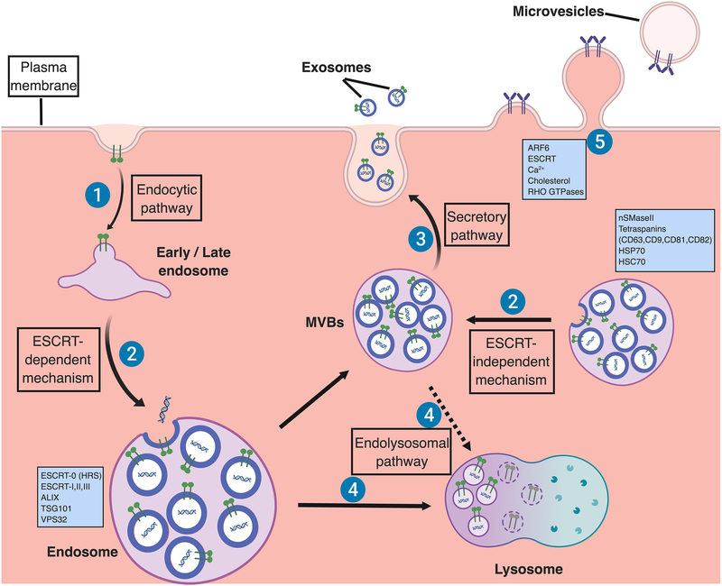
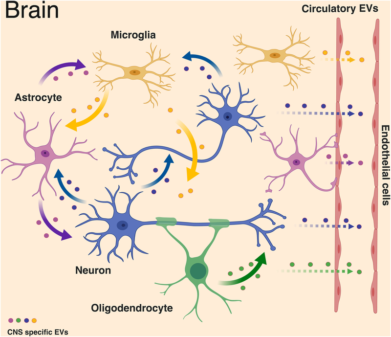
Extracellular vesicles (EVs) are heterogeneous cell-derived membranous vesicles which carry a large diversity of molecules such as proteins and RNA species. They are now considered to be a general mode of intercellular communication by direct transfer of biomolecules. Emerging evidence demonstrates that EVs are involved in multiple pathological processes of brain diseases including neurodegenerative disorders. In this review, we investigate the current knowledge about EV biology. We also provide an overview of the roles of EVs in related brain diseases, particularly in neurodegenerative disorders. Finally, we discuss their potential applications as novel biomarkers as well as the developments of EV-based therapies.

Keywords: Alzheimer’s disease; Biomarkers; Exosomes; Extracellular vesicles; Interleukins; Microtubule-associated protein tau; Neurodegenerative disorders.


Figures

Figure 1.
Figure 1.. Biogenesis of extracellular vesicles.
Exosomes are secreted from the multivesicular body (MVB), which is formed by invagination of the endosomal membrane. Initially, extracellular cargoes are targeted at the plasma membrane to form early endosomes by endocytic pathway (1). Early endosomes undergo transition to late endosomes and inward budding in which exosomal cargos including specific proteins and nucleic acids are further loaded to form multivesicular bodies (MVBs) containing intraluminal vesicles (ILVs) [11]. Endosomal sorting complex required for transport (ESCRT)-dependent mechanism, which are regulated by ESCRT proteins (ESCRT-0, I, II and III) and their accessories (ALIX, TSG101, VPS32) [66, 67], and ESCRT-independent mechanism, which are regulated by neutral sphingomyelinase 2 (nSMaseII), tetraspanins, and the chaperone heat shock proteins (HSP70, HSC70), can develop ILVs (2) [59, 70, 73, 83]. Some of MVBs can be further fused with the lysosomal membrane, resulting in the degradation of the ILVs and their contents for recycling as an endolysosomal pathway (3) [13, 14]. Alternatively, ILVs are released into the extracellular space as exosomes via a secretory pathway (4) [12]. For microvesicles, they are formed by direct outward budding of the plasma membrane, a process which is regulated by the ESCRT components and ADP ribosylation factor 6 (ARF6), some small GTPases, lipids, and Ca2+-dependent enzymatic machineries [1]. (Figure was created with BioRender. com)
Figure 2.
Figure 2.. Intercellular communication of neural cells derived extracellular vesicles in brain.
In the central nervous system, EVs could be secreted from one cell type and targeted to the others to engage multiple functions. Microglia have been proposed to secrete EVs containing the pro-inflammatory cytokine interleukin-1 β (IL-1 β), which mediates inflammasome response and the aminopeptidase CD13, which provides metabolic support [113, 198]. Similar to microglia, astrocyte-derived EVs play both neuroprotective and neurotoxic roles such as containing synapsin 1 to promote neurite outgrowth and Nef to mediate neurotoxicity [–117]. Oligodendrocytes secrete myelin molecules and stress-protective proteins in EVs, which are reported to participate in myelin formation and maintenance [118] as well as trophic support of neurons [8]. Neurodegenerative disorder-associated proteins such as prions [122], amyloid-β peptide [123], Tau [10], and α-synuclein [124] can also be released from EVs of the neural cells, leading to the spread of protein aggregate seeds and disease progression. In addition, these EVs could be exported through blood-brain barrier as circulatory EVs, which can be used for disease-specific biomarkers [1]. (Figure was created with BioRender. com)

References

    1. Thompson AG, Gray E, Heman-Ackah SM, Mager I, Talbot K, Andaloussi SE, et al. Extracellular vesicles in neurodegenerative disease - pathogenesis to biomarkers. Nat Rev Neurol. 2016;12(6):346–57. doi: 10.1038/nrneurol.2016.68. - DOI - PubMed
    1. Johnstone RM, Adam M, Hammond JR, Orr L, Turbide C. Vesicle formation during reticulocyte maturation. Association of plasma membrane activities with released vesicles (exosomes). J Biol Chem. 1987;262(19):9412–20. - PubMed
    1. Yanez-Mo M, Siljander PR, Andreu Z, Zavec AB, Borras FE, Buzas EI, et al. Biological properties of extracellular vesicles and their physiological functions. J Extracell Vesicles. 2015;4:27066. doi: 10.3402/jev.v4.27066. - DOI - PMC - PubMed
    1. Schorey JS, Cheng Y, Singh PP, Smith VL. Exosomes and other extracellular vesicles in host-pathogen interactions. EMBO Rep. 2015;16(1):24–43. doi: 10.15252/embr.201439363. - DOI - PMC - PubMed
    1. Kalra H, Simpson RJ, Ji H, Aikawa E, Altevogt P, Askenase P, et al. Vesiclepedia: a compendium for extracellular vesicles with continuous community annotation. PLoS Biol. 2012;10(12):e1001450. doi: 10.1371/journal.pbio.1001450. - DOI - PMC - PubMed

Publication types