

Postpartum ovarian vein thrombosis and venous anatomical variation
- PMID: 31229971
- PMCID: PMC6605948
- DOI: 10.1136/bcr-2018-228399
Postpartum ovarian vein thrombosis and venous anatomical variation
Abstract
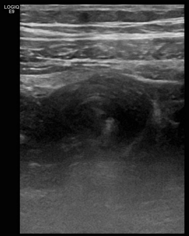
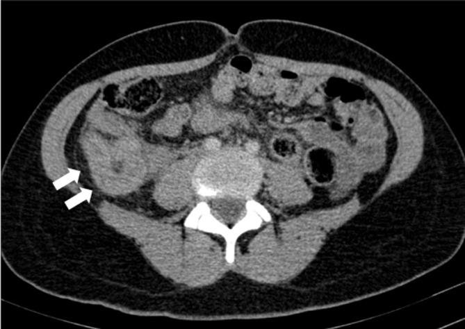
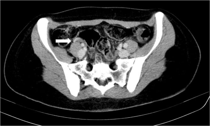
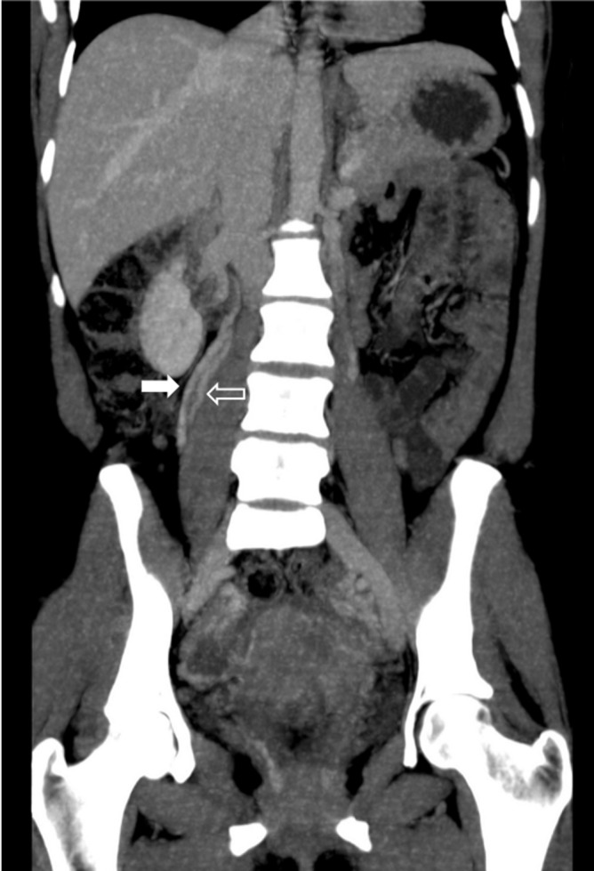
A 34-year-old multipara presented 72 hours postpartum with acute right-sided abdominal pain. The investigation revealed mild leucocytosis with positive D-dimer and elevated C reactive protein. Abdominal ultrasound and abdominopelvic CT demonstrated an enlarged right ovarian vein with endoluminal thrombus, representing postpartum ovarian vein thrombosis. The patient became asymptomatic 48 hours after starting broad-spectrum antibiotic treatment and anticoagulant therapy. She completed the treatment in ambulatory regimen and control abdominopelvic CT imaging was performed and revealed a duplicated right ovarian vein and a small residual subacute thrombus in the lumen of the distal right ovarian vein. The patient remained asymptomatic in the clinical follow-up.
Keywords: gynecology; pregnancy; radiology.
© BMJ Publishing Group Limited 2019. No commercial re-use. See rights and permissions. Published by BMJ.
Conflict of interest statement
Competing interests: None declared.
Figures
References
-
- Dougan C, Phillips R, Harley I, et al. Postpartum ovarian vein thrombosis. The Obstetrician & Gynaecologist 2016;18:291–9. 10.1111/tog.12295 - DOI
Publication types
MeSH terms
Substances
LinkOut - more resources
Full Text Sources
Medical
Research Materials