. 2020 Jan;36(1):1-10.
doi: 10.1007/s12264-019-00406-4. Epub 2019 Jun 22.

Inhibition of Muscular Nociceptive Afferents via the Activation of Cutaneous Nociceptors in a Rat Model of Inflammatory Muscle Pain

Affiliations

Inhibition of Muscular Nociceptive Afferents via the Activation of Cutaneous Nociceptors in a Rat Model of Inflammatory Muscle Pain

Yehong Fang et al. Neurosci Bull. 2020 Jan.

Abstract

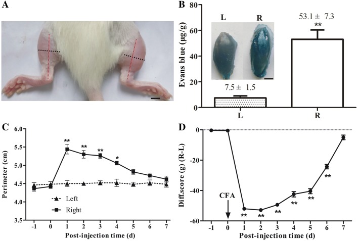
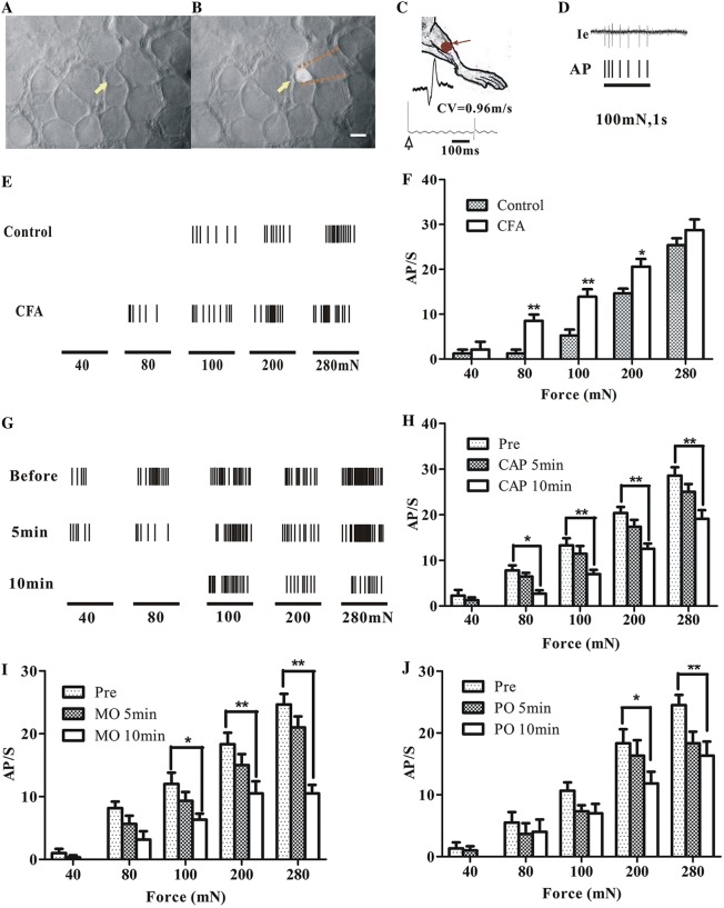
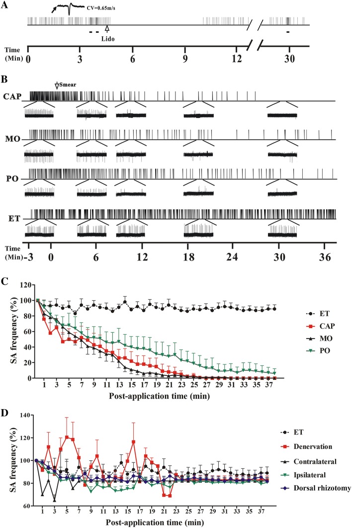
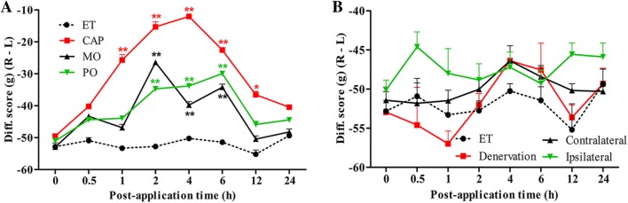
Topical irritants such as capsaicin (CAP), peppermint oil (PO), and mustard oil (MO) are effective in relieving inflammatory muscle pain. We investigated the effects of topical irritants in a rat model of inflammatory muscle pain produced by injecting complete Freund's adjuvant (CFA) into the tibialis anterior muscle. CFA-induced mechanical hypersensitivity and the spontaneous activity of muscular nociceptive afferents, and decreased weight-bearing of the hindlimb were relieved by topical application of CAP, PO, or MO on the skin overlying the inflamed muscle. The effects of topical irritants were abolished when applied to the skin on the ipsilateral plantar region or on the contralateral leg, or when the relevant cutaneous nerve or dorsal root was transected. Our results demonstrated that topical irritants may alleviate inflammatory muscle pain via activating cutaneous nociceptors and subsequently inhibiting the abnormal activity of muscular nociceptive neurons.

Keywords: Capsaicin; Cutaneous nociceptor; Inflammatory muscle pain; Muscular nociceptor.


Conflict of interest statement

All authors claim that there are no conflicts of interest.

Figures

Fig. 1
Fig. 1
Complete Freund’s adjuvant (CFA)-induced inflammatory muscle pain. A Representative photograph showing the swollen right hindlimb 2 days after CFA injection. Red and black dotted lines indicate the position of the tibia and the perimeter at the middle of the tibia, respectively. scale bar, 5 mm. B Evans blue extravasation from the ipsilateral (R) tibialis anterior 2 days after CFA injection; contralateral side (L) with injection of isotonic saline as control (n = 4). Inset, typical images of tibialis anterior muscles. Scale bar, 2 mm. C Perimeter of the ipsilateral hindlimb at the middle of the tibia peaked 1 day after CFA injection and then declined (n = 5). D Difference scores in weight-bearing (ipsilateral–contralateral) reached a minimum 1 day after CFA injection and was significantly reduced for 6 days (n = 6). *P < 0.05, **P < 0.01; ipsilateral vs contralateral in B and C, post-injection vs pre-injection −1 day in D.
Fig. 2
Fig. 2
Anterograde labelling of primary sensory neurons innervating tibialis anterior. A Immunofluorescent staining of L2–L6 dorsal root ganglia (DRGs) with the neuronal marker PGP9.5 showing primary sensory neurons in the L3–L5 DRGs innervating tibialis anterior anterogradely labeled with DiI (arrows). Inset, enlarged image of labeled neurons in L5 DRG. Scale bars, 100 μm (white); 20 μm (red). B Numbers of neurons labeled by DiI in L2–L6 DRGs (number of DRGs and rats in each group listed in brackets). C Numbers of large-(> 40 μm), medium (30 μm–40 μm), and small (< 30 μm) neurons labeled in L3–L5 DRGs. DiI, 1,1′-dioctadecyl-3,3,3′,3′-tetrame-thylindocarbocyanine perchlorate.
Fig. 3
Fig. 3
Mechanical thresholds of muscular C-nociceptive neurons in rat dorsal root ganglion (DRG). A Bright-field image of the surface of an L4 DRG; arrow indicates a small neuron. B Fluorescent image of the same cell body recorded by an extracellular glass micropipette (dotted lines). Scale bar, 20 μm. C Cartoon of measurement of conduction velocity of the sciatic nerve. Receptive field in tibialis anterior labeled by a red spot. D Single-neuron extracellular recording of a typical response of a fluorescence-labeled small neuron to 100 mN mechanical stimulation. Action potentials (APs) in the original recording trace (Ie) are presented as corresponding ticks below. E Responses of muscle nociceptive C-neurons to von Frey filaments of several forces (40 mN, 80 mN, 100 mN, 200 mN, and 280 mN) in CFA-injected and control rats. F Action potential discharge rates (AP/s) of muscular nociceptive C-neurons evoked by mechanical stimuli in complete Freund’s adjuvant (CFA)-injected (n = 11) and control rats (n = 6). G Typical recordings of the responses of a muscular C-nociceptive neuron from a CFA-injected rat to different forces applied to the receptive field in tibialis anterior before and after topical capsaicin (CAP) application. HJ Summary of responses (AP/s) of muscular C-nociceptive neurons to different forces before and after topical application of CAP (n = 11 in H), mustard oil (MO, n = 6 in I), and peppermint oil (PO, n = 6 in J) on the skin over tibialis anterior in CFA-injected rats. *P < 0.05, **P < 0.01; CFA vs control (F), post- vs pre-application (HJ).
Fig. 4
Fig. 4
Spontaneous activity (SA) of muscular nociceptive C-neurons recorded in L4 dorsal root ganglion (DRG) neurons after applying irritants in rats injected with complete Freund’s adjuvant (CFA). A Representative recording of SA in a muscular C-nociceptive neuron (conduction velocity [CV] = 0.65 m/s) 2 days after CFA injection. The SA was blocked within 1 min after intramuscular injection of lidocaine (Lido, 10 μL, blank arrow) into the receptive field, and then recovered ~6 min later. Horizontal bars below recording indicate mechanical stimuli (100 mN) to the receptive field. B Examples of SA in muscular nociceptive C-neurons that was gradually inhibited by capsaicin (CAP), mustard oil (MO), and peppermint oil (PO) on the skin overlying the inflamed muscle in CFA-injected rats. Ethanol (ET) was used as control. Insets below, expanded traces at different time points. C Statistics of SA frequency after applying CAP (n = 10), MO (n = 6), PO (n = 6), and ET (n = 8) on the skin overlying the inflamed muscle in CFA-injected rats. The average SA frequency within 3 min before irritant application was defined as 100%. CAP, MO and PO vs ET; CAP group: P < 0.05 at 5 min post-application and later; MO and PO groups: P < 0.05 at 12 min post-application and later. D Statistics of SA frequency after applying CAP to the skin overlying the inflamed muscle after cutaneous denervation (n = 6) or L4 dorsal rhizotomy (n = 2); the skin overlying the contralateral tibialis anterior (Contralateral) (n = 5); and the ipsilateral plantar skin (Ipsilateral) (n = 6) in CFA-injected rats. Denervation, L4 dorsal rhizotomy, Contralateral and Ipsilateral vs ET (n = 8); P > 0.05 at all tested time points.
Fig. 5
Fig. 5
Weight-bearing after application of topical irritants in CFA-injected rats. A Difference scores for weight-bearing (ipsilateral–contralateral) increased after the application of capsaicin (CAP) (n = 4), mustard oil (MO) (n = 4), peppermint oil (PO) (n = 4), and ethanol (ET) (n = 4) to the skin over the ipsilateral tibialis anterior. B Difference score for weight-bearing remained unchanged following the application of CAP to the denervated skin over the ipsilateral tibialis anterior (Denervation) (n = 7), skin over the contralateral tibialis anterior (Contralateral) (n = 6), and the ipsilateral plantar skin (Ipsilateral) (n = 6). ET on the skin overlying the inflamed muscle was taken as control in both A and B. *P < 0.05, **P < 0.01; CAP, MO and PO vs ET (A); Denervation, Contralateral and Ipsilateral vs ET (B).

Similar articles

Cited by

References

    1. Harden RN. Muscle pain syndromes. Am J Phys Med Rehabil. 2007;86:S47–S58. doi: 10.1097/PHM.0b013e31802ba648. - DOI - PubMed
    1. Staud R. Future perspectives: pathogenesis of chronic muscle pain. Best Pract Res Clin Rheumatol. 2007;21:581–596. doi: 10.1016/j.berh.2007.02.013. - DOI - PubMed
    1. Sumpton JE, Moulin DE. Fibromyalgia. Handb Clin Neurol. 2014;119:513–527. doi: 10.1016/B978-0-7020-4086-3.00033-3. - DOI - PubMed
    1. Borg-Stein J, Iaccarino MA. Myofascial pain syndrome treatments. Phys Med Rehabil Clin N Am. 2014;25:357–374. doi: 10.1016/j.pmr.2014.01.012. - DOI - PubMed
    1. Segal LS, Odgers R, Carpentieri D, Shrader MW. Back pain in Duchenne muscular dystrophy: steroids are not always the culprit. J Pediatr Orthop B. 2016;25:81–85. doi: 10.1097/BPB.0000000000000158. - DOI - PubMed

MeSH terms

LinkOut - more resources