. 2019 Aug;127(4):313-322.
doi: 10.1111/eos.12623. Epub 2019 Jun 22.

Selective bacterial degradation of the extracellular matrix attaching the gingiva to the tooth

Affiliations

Selective bacterial degradation of the extracellular matrix attaching the gingiva to the tooth

Aurélien Fouillen et al. Eur J Oral Sci. 2019 Aug.

Abstract

The junctional epithelium (JE) is a specialized portion of the gingiva that seals off the tooth-supporting tissues from the oral environment. This relationship is achieved via a unique adhesive extracellular matrix that is, in fact, a specialized basal lamina (sBL). Three unique proteins - amelotin (AMTN), odontogenic ameloblast-associated (ODAM), and secretory calcium-binding phosphoprotein proline-glutamine rich 1 (SCPPPQ1) - together with laminin-332 structure the supramolecular organization of this sBL and determine its adhesive capacity. Despite the constant challenge of the JE by the oral microbiome, little is known of the susceptibility of the sBL to bacterial degradation. Assays with trypsin-like proteases, as well as incubation with Porphyromonas gingivalis, Prevotella intermedia, and Treponema denticola, revealed that all constituents, except SCPPPQ1, were rapidly degraded. Porphyromonas gingivalis was also shown to alter the supramolecular network of reconstituted and native sBLs. These results provide evidence that proteolytic enzymes and selected gram-negative periodontopathogenic bacteria can attack this adhesive extracellular matrix, intimating that its degradation could contribute to progression of periodontal diseases.

Keywords: Porphyromonas gingivalis; junctional epithelium; periodontal diseases; specialized basal lamina; supramolecular network.


Conflict of interest statement

The authors declare no conflicts of interests.

Figures

Figure 1
Figure 1
Representative degradation profiles of amelotin (AMTN), odontogenic ameloblast‐associated (ODAM), and secretory calcium‐binding phosphoprotein proline‐glutamine rich 1 (SCPPPQ1) by periodontopathogenic bacteria. Western blot analysis following in‐vitro exposure to (A) Aggregatibacter actinomycetemcomitans and (B) Porphyromonas gingivalis. No degradation is observed with A. actinomycetemcomitans, whereas P. gingivalis degrades AMTN and ODAM in under 15 min, but SCPPPQ1 is not affected. Membranes were exposed for 1–2 s. MW in kDa.
Figure 2
Figure 2
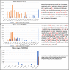
Top‐down mass spectrometry analysis of the cleavage products following in vitro incubation of proteins with Porphyromonas gingivalis. (A) Amelotin (AMTN), (B) odontogenic ameloblast‐associated (ODAM), and (C) secretory calcium‐binding phosphoprotein proline‐glutamine rich 1 (SCPPPQ1), before (blue) and after (orange) incubation with Porphyromonas gingivalis confirms the degradation of AMTN and ODAM and the non‐degradation of SCPPPQ1 observed by western blot analysis (Fig. 1 and S2). The right panels highlight the cleavage sites (red arrows) for the proteins.
Figure 3
Figure 3
Scanning electron microscopy of Porphyromonas gingivalis after 2 h of exposure to (A) buffer alone as the control and (B) buffer containing secretory calcium‐binding phosphoprotein proline‐glutamine rich 1 (SCPPPQ1). Following exposure to the protein, numerous outer membrane vesicles (OMVs) (arrows) appear on the bacterial surface.
Figure 4
Figure 4
SDSPAGE analysis of the in‐vitro digestion of (A) amelotin (AMTN), (B) odontogenic ameloblast‐associated (ODAM), and (C) secretory calcium‐binding phosphoprotein proline‐glutamine rich 1 (SCPPPQ1) with various enzymes reveals that the proteins are differentially susceptible to the various protease families tested. MW in kDa.
Figure 5
Figure 5
Atomic force microscopy (AFM) observation of degradation of reconstituted specialized basal lamina (sBL) by Porphyromonas gingivalis. (A) The supramolecular network formed by amelotin (AMTN), odontogenic ameloblast‐associated (ODAM), secretory calcium‐binding phosphoprotein proline‐glutamine rich 1 (SCPPPQ1), and laminin‐332 (Lm332), and (B, C) its local alteration (arrows) by P. gingivalis (Pg) to create a peribacterial region of destruction. (D, E) Scanning electron microscopy of the AFM preparations confirms the peripheral destruction and additionally shows the presence of residual filamentous material (*) in the affected region.
Figure 6
Figure 6
Scanning electron microscopy observations of the native specialized basal lamina (sBL) incubated with Porphyromonas gingivalis. (A) Overview (dashed area) and (B) higher‐magnification view of the exposed sBL on an enamel organ cap. Following exposure of the sBL to P. gingivalis for 2 h, (C) zones of alteration can be seen around the bacteria (white dashed lines); after 6 h (D) the peribacterial zones appear cavitated (arrow).
Figure 7
Figure 7
Scanning electron microscopy of junctional epithelium caps exposed to Porphyromonas gingivalis (A, B) for 6 h. Similarly to the exposure of enamel organ caps (Fig. 6), there are peribacterial alterations of the native specialized basal lamina (sBL) (arrows). Scale bar = 1 μm.

References

    1. Schroeder HE, Listgarten MA. The gingival tissues: the architecture of peridontal protection. Periodontol 2000 1997; 13: 91–120. - PubMed
    1. Bosshardt DD, Lang NP. The junctional epithelium: from health to disease. J Dent Res 2005; 84: 9–20. - PubMed
    1. Li X, Kolltveit KM, Tronstad L, Olsen I. Systemic diseases caused by oral infection. Clin Microbiol Rev 2000; 13: 547–558. - PMC - PubMed
    1. Hormia M, Owaribe K, Virtanen I. The dento‐epithelial junction: cell adhesion by type I hemidesmosomes in the absence of a true basal lamina. J Periodontol 2001; 72: 788–797. - PubMed
    1. Fouillen A, Dos Santos Neves J, Mary C, Castonguay JD, Moffatt P, Baron C, Nanci A. Interactions of AMTN, ODAM and SCPPPQ1 proteins of a specialized basal lamina that attaches epithelial cells to tooth mineral. Sci Rep 2017; 7: 46683. - PMC - PubMed

Publication types

Grants and funding

LinkOut - more resources