. 2019 Jul 3;105(1):132-150.
doi: 10.1016/j.ajhg.2019.05.015. Epub 2019 Jun 20.

The Genomics of Arthrogryposis, a Complex Trait: Candidate Genes and Further Evidence for Oligogenic Inheritance

Affiliations

The Genomics of Arthrogryposis, a Complex Trait: Candidate Genes and Further Evidence for Oligogenic Inheritance

Davut Pehlivan et al. Am J Hum Genet. .

Abstract

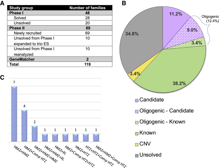
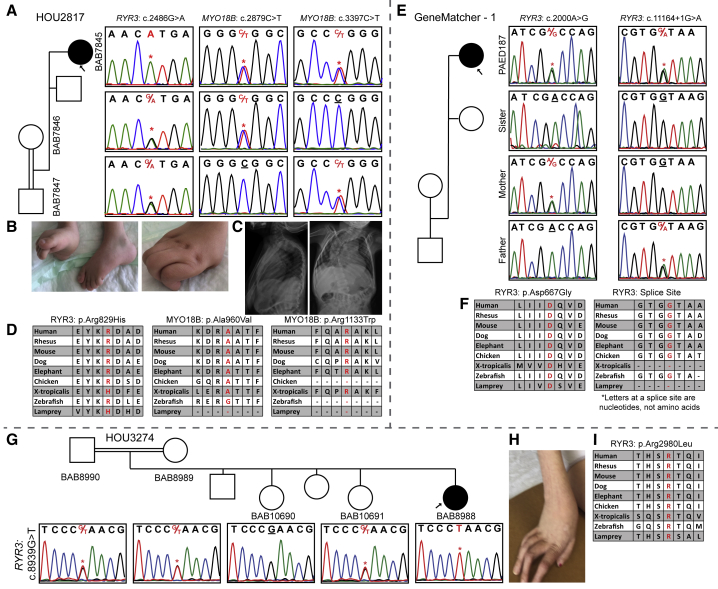
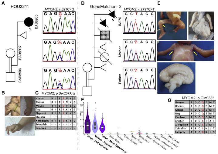
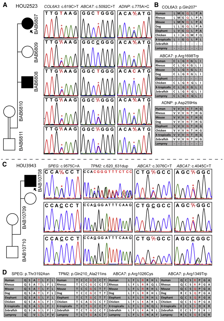
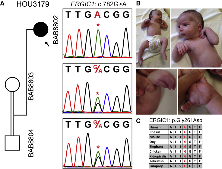
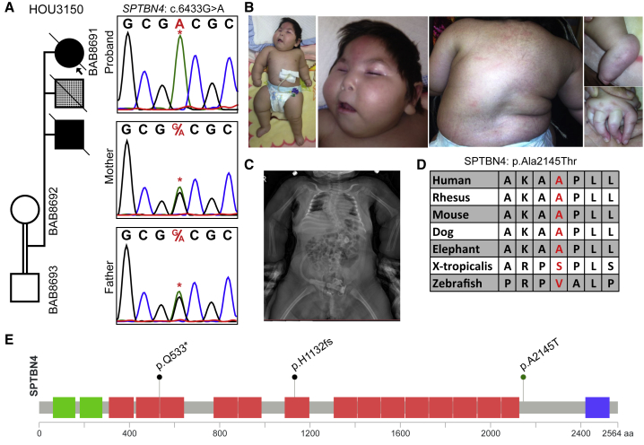
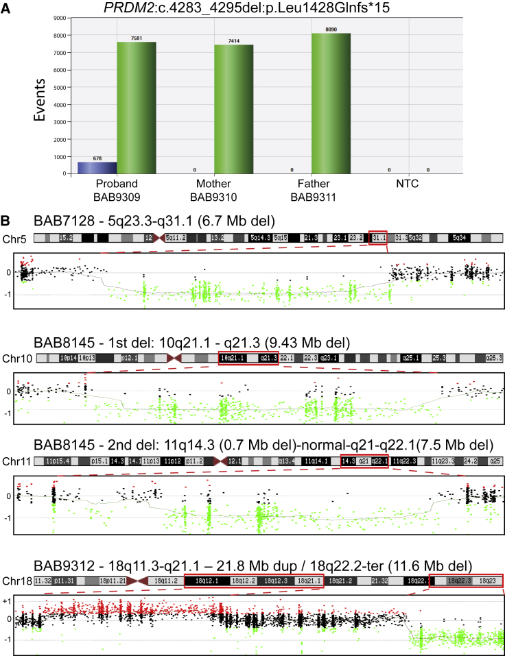
Arthrogryposis is a clinical finding that is present either as a feature of a neuromuscular condition or as part of a systemic disease in over 400 Mendelian conditions. The underlying molecular etiology remains largely unknown because of genetic and phenotypic heterogeneity. We applied exome sequencing (ES) in a cohort of 89 families with the clinical sign of arthrogryposis. Additional molecular techniques including array comparative genomic hybridization (aCGH) and Droplet Digital PCR (ddPCR) were performed on individuals who were found to have pathogenic copy number variants (CNVs) and mosaicism, respectively. A molecular diagnosis was established in 65.2% (58/89) of families. Eleven out of 58 families (19.0%) showed evidence for potential involvement of pathogenic variation at more than one locus, probably driven by absence of heterozygosity (AOH) burden due to identity-by-descent (IBD). RYR3, MYOM2, ERGIC1, SPTBN4, and ABCA7 represent genes, identified in two or more families, for which mutations are probably causative for arthrogryposis. We also provide evidence for the involvement of CNVs in the etiology of arthrogryposis and for the idea that both mono-allelic and bi-allelic variants in the same gene cause either similar or distinct syndromes. We were able to identify the molecular etiology in nine out of 20 families who underwent reanalysis. In summary, our data from family-based ES further delineate the molecular etiology of arthrogryposis, yielded several candidate disease-associated genes, and provide evidence for mutational burden in a biological pathway or network. Our study also highlights the importance of reanalysis of individuals with unsolved diagnoses in conjunction with sequencing extended family members.

Keywords: ES reanalysis; absence of heterozygosity; arthrogryposis; identity-by-descent; joint contracture; multilocus pathogenic variation; neuromuscular disease; trio-exome.


Figures

Figure 1
Figure 1
Distribution and Molecular Results of Enrolled Individuals in the Arthrogryposis Cohort and Families with Multilocus Pathogenic Variation (A) Distribution of individuals enrolled in arthrogryposis cohort. (B) A pie chart displaying the distribution of molecular findings for each individual in this study (i.e., phase II) of the arthrogryposis cohort (89 families). Multilocus pathogenic variant model families were classified according to muscle gene status. A corresponding explanation for the colors in the pie chart is below the chart. The abbreviation CNV = copy number variation. (C) Families affected with multilocus pathogenic variation (19 families) from phase I + II. Abbreviations are as follows: HMZ = homozygous, HTZ = heterozygous, Comp HTZ = compound heterozygous, and XL = hemizygous male.
Figure 2
Figure 2
Segregation Results, Images, and Variant Distributions of the Individuals with RYR3 Mutation (A) Sanger studies showing compound-heterozygous variants in MYO18B and a homozygous variant in RYR3 in the proband of family HOU2817. (B) Pictures of the proband’s contractures in fingers and toes. (C) Anterior-posterior and lateral views of spinal X-ray showing the kyphoscoliosis. (D) Conservation of the three variants through species. Variants of interest are written with a red font. (E) Segregation studies for both the c.2000A>G and c.11164+1G>A variants in GeneMatcher – 1. (F) Highly conserved asparagine (D) amino acid and splice site through species. (G) A pedigree and Sanger segregation of family HOU3274 that had a single affected female individual (BAB8988) and two unaffected siblings available for study. Both parents and one unaffected sibling are heterozygous, the second unaffected sibling is wild type, and our proband is homozygous for the c.8939G>T variant. (H) A colored picture of the hand showing the contractures in the fingers of individual BAB8988. (I) The arginine (R) amino acid residue is highly conserved among species and specified in red font.
Figure 3
Figure 3
Clinical and Molecular Details of the MYOM2 Individuals (A) Pedigree and segregation analyses of the identified MYOM2 variant (c.621C>G) in family HOU3211. The parents are shown as consanguineous because they are from the same small village and the proband demonstrates large AOH blocks on exome data. As expected with a recessive inheritance pattern, the parents are heterozygous and the proband (individual BAB8905) is homozygous for the c.621C>G variant. (B) Proband’s photos showing the contractures in the hands and feet. (C) High conservation of the Ser207 amino acid in other species. The serine (S) amino acid is written with a red font color. (D) A pedigree of the family (GeneMatcher – 2) showing a complicated medical history including affected and unaffected deceased siblings along with medically terminated or spontaneously aborted individuals in the GeneMatcher family. The fourth pregnancy is shown with a checkered box to indicate a different phenotype than neuromuscular disease (i.e., he was found to have cardiac and gastrointestinal anomalies along with intrauterine growth restriction). Sanger PCR from available individuals shows that the affected fetus is homozygous and the parents are heterozygous for the c.2797C>T variant. (E) Pictures of the fetus showing pterygia of axillae and popliteal joints and a midsagittal cut of fetal brain showing partial agenesis of the corpus callosum and bilateral hypoplastic lungs. (F) A genotype-tissue expression (GTEx) panel showing specific expression of MYOM2 in skeletal and heart muscles. (G) Amino acid conservation around Gln933 among other species. The Gln (Q) amino acid is written in red font.
Figure 4
Figure 4
Families with ABCA7 Variants (A) A pedigree suggesting an autosomal-recessive inheritance pattern for ABCA7, COL6A3, and ADNP in family HOU2523; the affected individuals are homozygous, whereas unaffected individuals are heterozygous. Individual BAB6807 has a homozygous stop-gain mutation in COL6A3 in addition to a homozygous ABCA7 variant shared with individual BAB6808. The ADNP variant is homozygous in individual BAB6808, who has a DD and ID phenotype. (B) Conservation profiles of all three genes. (C) A pedigree of family HOU3943 with segregation studies showing bi-allelic variants in ABCA7 and de novo heterozygous variants in SPEG and TPM2. (D) The amino acid alignment of the detected variants across different species.
Figure 5
Figure 5
Segregation, Pictures, and Protein Conservation for a Homozygous ERGIC1 Variant in Individual BAB8802 (A) Segregation analyses of the exome-detected variant; the proband was homozygous and the parents were heterozygous carriers, as expected in recessive disease traits. (B) Proband photographs showing restrictions in the wrists and fingers and the pes equinovarus deformity in the feet. (C) A peptide alignment showing the conservation of the affected amino acid across species.
Figure 6
Figure 6
Clinical and Molecular Studies for a Homozygous SPTBN4 Variant in Individual BAB8691 (A) Segregation studies showing heterozygosity for the parents and homozygosity for the index for variant c.6433G>A. The second child, who died of an unknown lung malformation, is shown with a checkered box. (B) Pictures of the proband showing a myopathic face, scoliosis, contractures in the fingers, and pes equinovarus deformity in the feet. (C) An anterior-posterior view of a spine X-ray showing severe thoracic scoliosis. (D) High conservation of the mutated amino acid residue across species (red font). (E) Protein domains of SPTBN4, with the variant identified in the present cohort (p.Ala2145Thr) and variants reported in two individuals in the literature (p.Gln533 and p.His1132fs).
Figure 7
Figure 7
Molecular Studies in Individual BAB9309 and Copy Number Variant Families (A) Droplet digital PCR (ddPCR) results for the family; the results show 8% mosaicism for the PRDM2 variant in the proband, and this finding is consistent with the exome mosaicism rate (8%) and no deletion in the healthy parents. (B) Results for aCGH analyses in three individuals are shown. Each dot indicates oligonucleotide probes: black dots represent a normal copy number, red dots represent a copy number gain, and green dots represent copy number losses as compared with a gender-matched control. The proband’s designated ID (BAB#s), the chromosomal location, and the approximate sizes of the copy number variants (CNVs) are written above each chromosome ideogram, and the chromosome number is written on the left side of ideogram. Red dashed lines determine the chromosomal region investigated by aCGH. Abbreviations are as follows: Chr = chromosome, Del = deletion, Dup = duplication, Mb = megabase, NTC = no template control, and Ter = terminal.

Similar articles

  • High prevalence of multilocus pathogenic variation in neurodevelopmental disorders in the Turkish population.
    Mitani T, Isikay S, Gezdirici A, Gulec EY, Punetha J, Fatih JM, Herman I, Akay G, Du H, Calame DG, Ayaz A, Tos T, Yesil G, Aydin H, Geckinli B, Elcioglu N, Candan S, Sezer O, Erdem HB, Gul D, Demiral E, Elmas M, Yesilbas O, Kilic B, Gungor S, Ceylan AC, Bozdogan S, Ozalp O, Cicek S, Aslan H, Yalcintepe S, Topcu V, Bayram Y, Grochowski CM, Jolly A, Dawood M, Duan R, Jhangiani SN, Doddapaneni H, Hu J, Muzny DM; Baylor-Hopkins Center for Mendelian Genomics; Marafi D, Akdemir ZC, Karaca E, Carvalho CMB, Gibbs RA, Posey JE, Lupski JR, Pehlivan D. Mitani T, et al. Am J Hum Genet. 2021 Oct 7;108(10):1981-2005. doi: 10.1016/j.ajhg.2021.08.009. Epub 2021 Sep 28. Am J Hum Genet. 2021. PMID: 34582790 Free PMC article.
  • Bi-allelic loss of ERGIC1 causes relatively mild arthrogryposis.
    Marconi C, Lemmens L, Masclaux F, Mattioli F, Fluss J, Extermann P, Mendez P, Leuchter RH, Stathaki E, Laurent S, Hammar E, Vannier A, Varvagiannis K, Guipponi M, Sloan-Bena F, Blouin JL, Abramowicz M, Fokstuen S. Marconi C, et al. Clin Genet. 2021 Sep;100(3):329-333. doi: 10.1111/cge.14004. Epub 2021 Jun 14. Clin Genet. 2021. PMID: 34037256 Free PMC article.
  • The genomic and clinical landscape of fetal akinesia.
    Pergande M, Motameny S, Özdemir Ö, Kreutzer M, Wang H, Daimagüler HS, Becker K, Karakaya M, Ehrhardt H, Elcioglu N, Ostojic S, Chao CM, Kawalia A, Duman Ö, Koy A, Hahn A, Reimann J, Schoner K, Schänzer A, Westhoff JH, Schwaibold EMC, Cossee M, Imbert-Bouteille M, von Pein H, Haliloglu G, Topaloglu H, Altmüller J, Nürnberg P, Thiele H, Heller R, Cirak S. Pergande M, et al. Genet Med. 2020 Mar;22(3):511-523. doi: 10.1038/s41436-019-0680-1. Epub 2019 Nov 4. Genet Med. 2020. PMID: 31680123
  • Insights into genetics, human biology and disease gleaned from family based genomic studies.
    Posey JE, O'Donnell-Luria AH, Chong JX, Harel T, Jhangiani SN, Coban Akdemir ZH, Buyske S, Pehlivan D, Carvalho CMB, Baxter S, Sobreira N, Liu P, Wu N, Rosenfeld JA, Kumar S, Avramopoulos D, White JJ, Doheny KF, Witmer PD, Boehm C, Sutton VR, Muzny DM, Boerwinkle E, Günel M, Nickerson DA, Mane S, MacArthur DG, Gibbs RA, Hamosh A, Lifton RP, Matise TC, Rehm HL, Gerstein M, Bamshad MJ, Valle D, Lupski JR; Centers for Mendelian Genomics. Posey JE, et al. Genet Med. 2019 Apr;21(4):798-812. doi: 10.1038/s41436-018-0408-7. Epub 2019 Jan 18. Genet Med. 2019. PMID: 30655598 Free PMC article. Review.
  • Familial Gordon syndrome associated with a PIEZO2 mutation.
    Alisch F, Weichert A, Kalache K, Paradiso V, Longardt AC, Dame C, Hoffmann K, Horn D. Alisch F, et al. Am J Med Genet A. 2017 Jan;173(1):254-259. doi: 10.1002/ajmg.a.37997. Epub 2016 Oct 7. Am J Med Genet A. 2017. PMID: 27714920 Review.

Cited by

  • Phenotypic spectrum and genomics of undiagnosed arthrogryposis multiplex congenita.
    Laquerriere A, Jaber D, Abiusi E, Maluenda J, Mejlachowicz D, Vivanti A, Dieterich K, Stoeva R, Quevarec L, Nolent F, Biancalana V, Latour P, Sternberg D, Capri Y, Verloes A, Bessieres B, Loeuillet L, Attie-Bitach T, Martinovic J, Blesson S, Petit F, Beneteau C, Whalen S, Marguet F, Bouligand J, Héron D, Viot G, Amiel J, Amram D, Bellesme C, Bucourt M, Faivre L, Jouk PS, Khung S, Sigaudy S, Delezoide AL, Goldenberg A, Jacquemont ML, Lambert L, Layet V, Lyonnet S, Munnich A, Van Maldergem L, Piard J, Guimiot F, Landrieu P, Letard P, Pelluard F, Perrin L, Saint-Frison MH, Topaloglu H, Trestard L, Vincent-Delorme C, Amthor H, Barnerias C, Benachi A, Bieth E, Boucher E, Cormier-Daire V, Delahaye-Duriez A, Desguerre I, Eymard B, Francannet C, Grotto S, Lacombe D, Laffargue F, Legendre M, Martin-Coignard D, Mégarbané A, Mercier S, Nizon M, Rigonnot L, Prieur F, Quélin C, Ranjatoelina-Randrianaivo H, Resta N, Toutain A, Verhelst H, Vincent M, Colin E, Fallet-Bianco C, Granier M, Grigorescu R, Saada J, Gonzales M, Guiochon-Mantel A, Bessereau JL, Tawk M, Gut I, Gitiaux C, Melki J. Laquerriere A, et al. J Med Genet. 2022 Jun;59(6):559-567. doi: 10.1136/jmedgenet-2020-107595. Epub 2021 Apr 5. J Med Genet. 2022. PMID: 33820833 Free PMC article.
  • Quantitative dissection of multilocus pathogenic variation in an Egyptian infant with severe neurodevelopmental disorder resulting from multiple molecular diagnoses.
    Herman I, Jolly A, Du H, Dawood M, Abdel-Salam GMH, Marafi D, Mitani T, Calame DG, Coban-Akdemir Z, Fatih JM, Hegazy I, Jhangiani SN, Gibbs RA, Pehlivan D, Posey JE, Lupski JR. Herman I, et al. Am J Med Genet A. 2022 Mar;188(3):735-750. doi: 10.1002/ajmg.a.62565. Epub 2021 Nov 23. Am J Med Genet A. 2022. PMID: 34816580 Free PMC article.
  • Is Gene-Size an Issue for the Diagnosis of Skeletal Muscle Disorders?
    Savarese M, Välipakka S, Johari M, Hackman P, Udd B. Savarese M, et al. J Neuromuscul Dis. 2020;7(3):203-216. doi: 10.3233/JND-190459. J Neuromuscul Dis. 2020. PMID: 32176652 Free PMC article. Review.
  • The mutational burden and oligogenic inheritance in Klippel-Feil syndrome.
    Li Z, Zhao S, Cai S, Zhang Y, Wang L, Niu Y, Li X, Hu J, Chen J, Wang S, Wang H, Liu G, Tian Y, Wu Z, Zhang TJ; DISCO (Deciphering Disorders Involving Scoliosis and COmorbidities) study; Wang Y, Wu N. Li Z, et al. BMC Musculoskelet Disord. 2020 Apr 11;21(1):220. doi: 10.1186/s12891-020-03229-x. BMC Musculoskelet Disord. 2020. PMID: 32278351 Free PMC article.
  • Mechanisms of node of Ranvier assembly.
    Rasband MN, Peles E. Rasband MN, et al. Nat Rev Neurosci. 2021 Jan;22(1):7-20. doi: 10.1038/s41583-020-00406-8. Epub 2020 Nov 25. Nat Rev Neurosci. 2021. PMID: 33239761 Review.

References

    1. Hall J.G. Arthrogryposis (multiple congenital contractures): Diagnostic approach to etiology, classification, genetics, and general principles. Eur. J. Med. Genet. 2014;57:464–472. - PubMed
    2. Hall, J.G. (2014). Arthrogryposis (multiple congenital contractures): Diagnostic approach to etiology, classification, genetics, and general principles. Eur. J. Med. Genet. 57, 464-472. - PubMed
    1. Lowry R.B., Sibbald B., Bedard T., Hall J.G. Prevalence of multiple congenital contractures including arthrogryposis multiplex congenita in Alberta, Canada, and a strategy for classification and coding. Birth Defects Res. A Clin. Mol. Teratol. 2010;88:1057–1061. - PubMed
    2. Lowry, R.B., Sibbald, B., Bedard, T., and Hall, J.G. (2010). Prevalence of multiple congenital contractures including arthrogryposis multiplex congenita in Alberta, Canada, and a strategy for classification and coding. Birth Defects Res. A Clin. Mol. Teratol. 88, 1057-1061. - PubMed
    1. Hall J.G., Kiefer J. Arthrogryposis as a syndrome: Gene ontology analysis. Mol. Syndromol. 2016;7:101–109. - PMC - PubMed
    2. Hall, J.G., and Kiefer, J. (2016). Arthrogryposis as a syndrome: Gene ontology analysis. Mol. Syndromol. 7, 101-109. - PMC - PubMed
    1. Balta B., Erdogan M., Ergul A.B., Sahin Y., Ozcan A. Interstitial deletion 5p14.1-p15.2 and 5q14.3-q23.2 in a patient with clubfoot, blepharophimosis, arthrogryposis, and multiple congenital abnormalities. Am. J. Med. Genet. A. 2017;173:2798–2802. - PubMed
    2. Balta, B., Erdogan, M., Ergul, A.B., Sahin, Y., and Ozcan, A. (2017). Interstitial deletion 5p14.1-p15.2 and 5q14.3-q23.2 in a patient with clubfoot, blepharophimosis, arthrogryposis, and multiple congenital abnormalities. Am. J. Med. Genet. A. 173, 2798-2802. - PubMed
    1. Carrascosa-Romero M.C., Suela J., Pardal-Fernández J.M., Bermejo-Sánchez E., Vidal-Company A., MacDonald A., Tébar-Gil R., Martínez-Fernández M.L., Martínez-Frías M.L. A 2.84 Mb deletion at 21q22.11 in a patient clinically diagnosed with Marden-Walker syndrome. Am. J. Med. Genet. A. 2013;161A:2281–2290. - PubMed
    2. Carrascosa-Romero, M.C., Suela, J., Pardal-Fernandez, J.M., Bermejo-Sanchez, E., Vidal-Company, A., MacDonald, A., Tebar-Gil, R., Martinez-Fernandez, M.L., and Martinez-Frias, M.L. (2013). A 2.84 Mb deletion at 21q22.11 in a patient clinically diagnosed with Marden-Walker syndrome. Am. J. Med. Genet. A. 161A, 2281-2290. - PubMed

Publication types

MeSH terms