. 2019 Jul 3;105(1):213-220.
doi: 10.1016/j.ajhg.2019.05.005. Epub 2019 Jun 20.

De Novo Variants in TAOK1 Cause Neurodevelopmental Disorders

Affiliations

De Novo Variants in TAOK1 Cause Neurodevelopmental Disorders

Marija Dulovic-Mahlow et al. Am J Hum Genet. .

Abstract

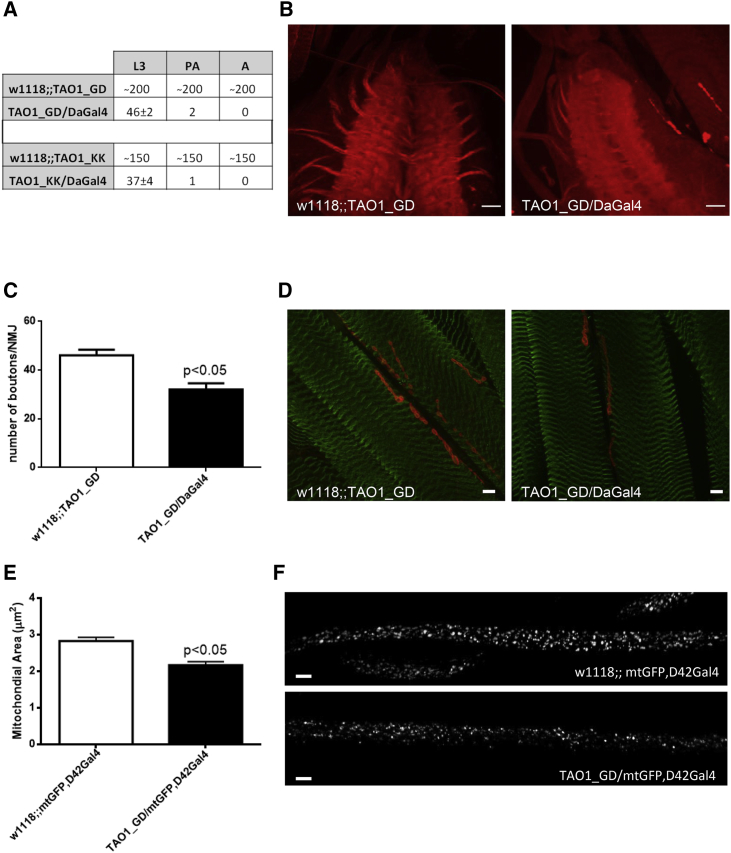
De novo variants represent a significant cause of neurodevelopmental delay and intellectual disability. A genetic basis can be identified in only half of individuals who have neurodevelopmental disorders (NDDs); this indicates that additional causes need to be elucidated. We compared the frequency of de novo variants in patient-parent trios with (n = 2,030) versus without (n = 2,755) NDDs. We identified de novo variants in TAOK1 (thousand and one [TAO] amino acid kinase 1), which encodes the serine/threonine-protein kinase TAO1, in three individuals with NDDs but not in persons who did not have NDDs. Through further screening and the use of GeneMatcher, five additional individuals with NDDs were found to have de novo variants. All eight variants were absent from gnomAD (Genome Aggregation Database). The variant carriers shared a non-specific phenotype of developmental delay, and six individuals had additional muscular hypotonia. We established a fibroblast line of one mutation carrier, and we demonstrated that reduced mRNA levels of TAOK1 could be increased upon cycloheximide treatment. These results indicate nonsense-mediated mRNA decay. Further, there was neither detectable phosphorylated TAO1 kinase nor phosphorylated tau in these cells, and mitochondrial morphology was altered. Knockdown of the ortholog gene Tao1 (Tao, CG14217) in Drosophila resulted in delayed early development. The majority of the Tao1-knockdown flies did not survive beyond the third instar larval stage. When compared to control flies, Tao1 knockdown flies revealed changed morphology of the ventral nerve cord and the neuromuscular junctions as well as a decreased number of endings (boutons). Furthermore, mitochondria in mutant flies showed altered distribution and decreased size in axons of motor neurons. Thus, we provide compelling evidence that de novo variants in TAOK1 cause NDDs.

Keywords: TAO kinase 1; de novo variants; fly model; neurodevelopmental disorders.


Figures

Figure 1
Figure 1
De novo Variants in TAOK1 Detected in Individuals with NDDs (A) Photographs of individual 6 show dysmorphic features including microsomia with short proximal extremities, macrocephaly with a high forehead, low-set ears, and down-slanting eyelids. The asymmetry of the lips is caused by the surgical intervention to correct the cleft lip and is therefore a secondary phenomenon. (B) Photographs of individual 8 show dysmorphic features including a long face, a high forehead, big ears, a long nose, down-slanting palpebral fissures, retrognathia, congenital flat feet, and hyperextensibility of the distal joints. The latter three features might be linked to the FBNI mutation and fit the diagnosis of Marfan syndrome. (C) Schematic representation of the encoded TAO1 kinase with its 1,001 amino acids. The functional domains (protein kinase [amino acids 28–281] and coiled-coil motifs 1 [amino acids 458–651] and 2 [amino acids 754–877]) are highlighted in red and blue, respectively. Arrows indicate the locations of the detected variants (see Table 1 for further descriptions). GenBank: NM_020791.2; NP_065842.1.
Figure 2
Figure 2
Characterization of the TAOK1 Variant c.2366_2367insC in Blood and a Fibroblast Line Derived from the Mutation Carrier (A) Electropherograms of Sanger sequencing of genomic (gDNA) and complementary DNA (cDNA) of individual 7 in the reverse direction. The variant is shown on the genomic level (upper left panel) from a blood sample, and the inserted C (G on the reverse strand) is highlighted. The expression of the mutant allele is decreased in blood (lower left panel), and in the fibroblast line (upper right panel) compared to the wild-type allele, as shown by the lower height of the peaks using Sanger sequencing. The mutant allele could be stabilized upon treatment with cycloheximide (CHX, 100 μg/mL, 8 h) as illustrated in the lower right panel. (B) The quantification of TAOK1 mRNA levels by real-time PCR indicated highly reduced levels of TAOK1 in the affected individual (AI) compared to the control individuals (controls) in blood and fibroblasts. The expression in fibroblasts can be increased upon treatment with cycloheximide (CHX). (C) Immunoblot analysis of total protein extract from fibroblasts of the affected individual (AI) and a control (control 1) with an antibody against phosphorylated TAO kinases TAO1, TAO2, and TAO3 indicates the absence of detectable, phosphorylated TAO1 kinase (p-TAOK1) in cells of the affected individual. Phosphorylation and abundance of TAO2 and 3 kinases (p-TAOK2/3) seemed to be unaltered. β-actin (ACTIN) was used as a loading control. (D) Immunoblot analysis of total protein extract from the fibroblast line of the affected individual (AI) and two controls (control 1, control 2) with antibodies against total TAOK1 and phosphorylated tau protein (p-tau) indicating absence of both TAOK1 and p-tau in cells of the affected individual. β-actin (ACTN) was used as a loading control. (E) Form factor as a measurement for mitochondrial interconnectivity was calculated in the fibroblast line of the affected individual (AI) and two controls (control 1, control 2) (10 cells each). Each dot represents the value in a single cell, and the mean and the interquartile range per individual are indicated.
Figure 3
Figure 3
Impaired Neurodevelopment in Drosophila with Knockdown of Tao1 (A) Early lethality in Tao1 knockdown flies. The table indicates the number of living flies in different stages (L3 — third instar larvae stage; PA — pharate adult stage; A — adult flies). Two Tao1 RNAi lines (TAO1_GD and TAO1_KK) were crossed with w1118 control line and the DaGal4 driver, which ubiquitously knocks down Tao1. (B) Smaller-sized ventral nerve cord in Tao1 knockdown flies. Representative images of L3 control (w1118;;TAO1_GD) and L3 Tao1 knockdown (TAO1_GD/DaGal4) larval ventral nerve cord (VNC) are shown, indicating a reduction in the size of VNC and less-developed axons starting from VNC in Tao1 knockdown flies. Immunolabeling of the L3 larval VNC was done with the synaptic marker anti-DLG (red). Four animals per genotype were analyzed. The scale bar represents 20 μm. (C) Significantly decreased amount of neuromuscular junction endings (boutons) in Tao1 knockdown flies. Quantification of boutons in control (w1118;;TAO1_GD) and Tao1 knockdown (TAO1_GD/DaGal4) larvae. p < 0.05 — significantly decreased number of boutons compared to those of the control; 15 images from four animals per genotype were analyzed. Error bars represent the SEM. (D) Representative images of the neuromuscular junctions and boutons are shown, indicating changed morphology and a reduced number of boutons. Immunolabeling of L3 control (w1118;;TAO1_GD) and L3 Tao1 knockdown (TAO1_GD/DaGal4) larval neuromuscular junctions (NMJs) and their boutons with the synaptic marker anti-DLG (red) and anti-GABARAB (green) defined muscular structure. The scale bar represents 10 μm. (E) Significantly decreased average mitochondrial size (μm2) in axons of Tao1 knockdown flies. The control line (w1118) and Tao1 RNAi line (TAO1_GD) were crossed with flies expressing mitochondrial-tagged GFP, predominantly in the motor neurons (mtGFP,D42Gal4), and quantification of axonal mitochondrial area in control (w1118;;mtGFP,D42Gal4) and Tao1 knockdown (TAO1_GD/mtGFP,D42Gal4) L3 larvae is shown. p < 0.05 — significantly decreased average mitochondrial area compared to the control; 10 images from three animals per genotype were analyzed. Error bars represent the SEM. (F) Representative images of axonal mitochondria are shown, indicating disturbed mitochondrial distribution and reduced mitochondrial size. The scale bar represents 10 μm.

References

    1. Sheridan E., Wright J., Small N., Corry P.C., Oddie S., Whibley C., Petherick E.S., Malik T., Pawson N., McKinney P.A., Parslow R.C. Risk factors for congenital anomaly in a multiethnic birth cohort: an analysis of the Born in Bradford study. Lancet. 2013;382:1350–1359. - PubMed
    2. Sheridan, E., Wright, J., Small, N., Corry, P.C., Oddie, S., Whibley, C., Petherick, E.S., Malik, T., Pawson, N., McKinney, P.A., and Parslow, R.C. (2013). Risk factors for congenital anomaly in a multiethnic birth cohort: an analysis of the Born in Bradford study. Lancet 382, 1350-1359. - PubMed
    1. Hoischen A., Krumm N., Eichler E.E. Prioritization of neurodevelopmental disease genes by discovery of new mutations. Nat. Neurosci. 2014;17:764–772. - PMC - PubMed
    2. Hoischen, A., Krumm, N., and Eichler, E.E. (2014). Prioritization of neurodevelopmental disease genes by discovery of new mutations. Nat. Neurosci. 17, 764-772. - PMC - PubMed
    1. Ku C.S., Polychronakos C., Tan E.K., Naidoo N., Pawitan Y., Roukos D.H., Mort M., Cooper D.N. A new paradigm emerges from the study of de novo mutations in the context of neurodevelopmental disease. Mol. Psychiatry. 2013;18:141–153. - PubMed
    2. Ku, C.S., Polychronakos, C., Tan, E.K., Naidoo, N., Pawitan, Y., Roukos, D.H., Mort, M., and Cooper, D.N. (2013). A new paradigm emerges from the study of de novo mutations in the context of neurodevelopmental disease. Mol. Psychiatry 18, 141-153. - PubMed
    1. Wright C.F., McRae J.F., Clayton S., Gallone G., Aitken S., FitzGerald T.W., Jones P., Prigmore E., Rajan D., Lord J., DDD Study Making new genetic diagnoses with old data: iterative reanalysis and reporting from genome-wide data in 1,133 families with developmental disorders. Genet. Med. 2018;20:1216–1223. - PMC - PubMed
    2. Wright, C.F., McRae, J.F., Clayton, S., Gallone, G., Aitken, S., FitzGerald, T.W., Jones, P., Prigmore, E., Rajan, D., Lord, J., et al.; DDD Study (2018). Making new genetic diagnoses with old data: iterative reanalysis and reporting from genome-wide data in 1,133 families with developmental disorders. Genet. Med. 20, 1216-1223. - PMC - PubMed
    1. Aronson S.J., Rehm H.L. Building the foundation for genomics in precision medicine. Nature. 2015;526:336–342. - PMC - PubMed
    2. Aronson, S.J., and Rehm, H.L. (2015). Building the foundation for genomics in precision medicine. Nature 526, 336-342. - PMC - PubMed

Publication types

Substances