

Novel Object Recognition in Rats With NMDAR Dysfunction in CA1 After Stereotactic Injection of Anti-NMDAR Encephalitis Cerebrospinal Fluid
- PMID: 31231304
- PMCID: PMC6560222
- DOI: 10.3389/fneur.2019.00586
Novel Object Recognition in Rats With NMDAR Dysfunction in CA1 After Stereotactic Injection of Anti-NMDAR Encephalitis Cerebrospinal Fluid
Abstract
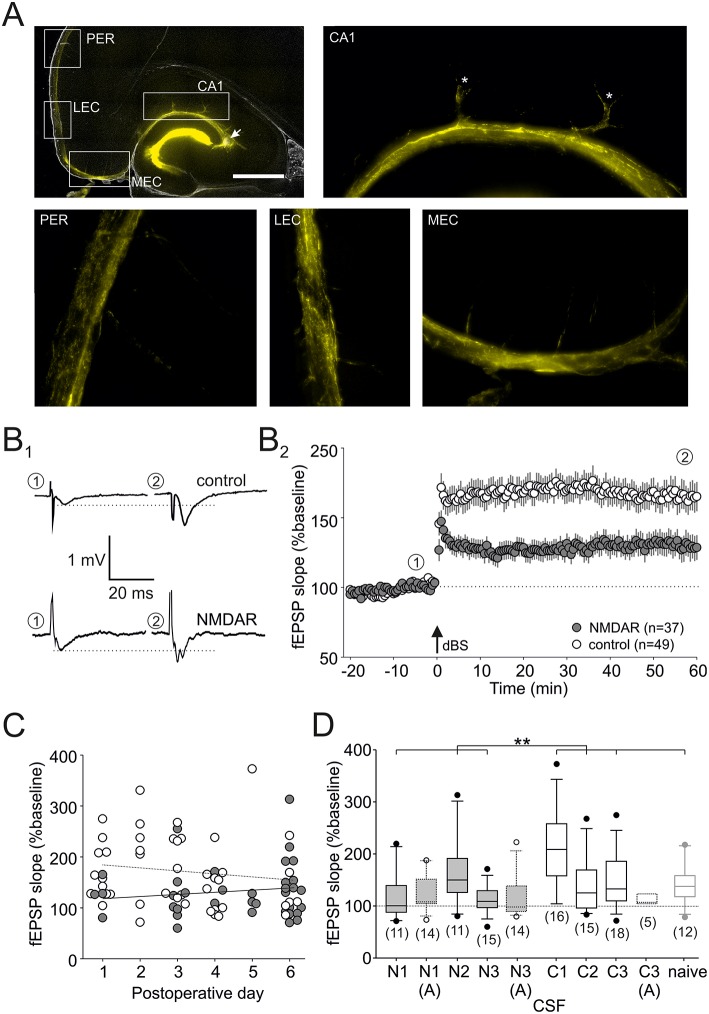
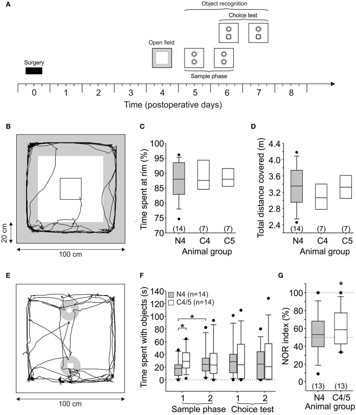
Purpose: Limbic encephalitis associated with autoantibodies against N-methyl D-aspartate receptors (NMDARs) often presents with memory impairment. NMDARs are key targets for memory acquisition and retrieval, and have been mechanistically linked to its underlying process, synaptic plasticity. Clinically, memory deficits are largely compatible with a pre-dominantly hippocampus-dependent phenotype, which, in rodents, is principally involved in spatial memory. Previous studies confirmed the impaired spatial memory in the rat model of anti-NMDAR encephalitis. Here, we hypothesized that non-spatial memory functions, such as object recognition might also be affected in this model. Methods: We performed stereotactic intrahippocampal bolus injection of human cerebrospinal fluid (CSF) from anti-NMDAR encephalitis and control patients into the hippocampus of the anesthetized rat. After recovery for 1-8 days, hippocampal slices were prepared from these animals and NMDAR-dependent long-term potentiation was assessed at the Schaffer collateral-CA1 synapse. In addition, we performed behavioral analyses using the open field and novel object recognition tasks. Results: NMDAR-dependent long-term potentiation in the hippocampal CA1 area was significantly suppressed, indicating successful NMDAR dysfunction in this subfield. Spontaneous locomotor activity as well as anxiety-related behavior in the open field did not differ between NMDAR-CSF-treated and control animals. In the novel object recognition task, there were no differences in the motivation to approach objects. In contrast, we observed a significantly preferred exploration of the novel object only in control, but not in NMDAR-CSF-treated rats. Conclusion: These results indicate that NMDAR dysfunction obtained by intrahippocampal stereotactic injection does not alter locomotor or anxiety-related behavior. In addition, approach to an object or exploratory behavior in general are not affected either, but intact initial NMDAR-dependent processes might be involved in novel object recognition.
Keywords: anti-NMDAR encephalitis; cerebrospinal fluid; long-term potentiation; object recognition; perirhinal cortex.
Figures
Similar articles
-
Differentially Altered NMDAR Dependent and Independent Long-Term Potentiation in the CA3 Subfield in a Model of Anti-NMDAR Encephalitis.Front Synaptic Neurosci. 2018 Jul 31;10:26. doi: 10.3389/fnsyn.2018.00026. eCollection 2018. Front Synaptic Neurosci. 2018. PMID: 30108497 Free PMC article.
-
Suppression of synaptic plasticity by cerebrospinal fluid from anti-NMDA receptor encephalitis patients.Neurobiol Dis. 2012 Jan;45(1):610-5. doi: 10.1016/j.nbd.2011.09.019. Epub 2011 Oct 8. Neurobiol Dis. 2012. PMID: 22008231
-
Ephrin-B2 prevents N-methyl-D-aspartate receptor antibody effects on memory and neuroplasticity.Ann Neurol. 2016 Sep;80(3):388-400. doi: 10.1002/ana.24721. Epub 2016 Aug 2. Ann Neurol. 2016. PMID: 27399303 Free PMC article.
-
Long-term potentiation and the role of N-methyl-D-aspartate receptors.Brain Res. 2015 Sep 24;1621:5-16. doi: 10.1016/j.brainres.2015.01.016. Epub 2015 Jan 22. Brain Res. 2015. PMID: 25619552 Free PMC article. Review.
-
Hippocampal NMDA receptors and the previous experience effect on memory.J Physiol Paris. 2014 Sep-Dec;108(4-6):263-9. doi: 10.1016/j.jphysparis.2014.08.001. Epub 2014 Aug 15. J Physiol Paris. 2014. PMID: 25132342 Review.
Cited by
-
Antidepressant Effects of (S)-Ketamine through a Reduction of Hyperpolarization-Activated Current Ih.iScience. 2020 Jun 26;23(6):101239. doi: 10.1016/j.isci.2020.101239. Epub 2020 Jun 4. iScience. 2020. PMID: 32629607 Free PMC article.
-
A peptide from the Japanese encephalitis virus failed to induce the production of anti-N-methyl-d-aspartate receptor antibodies via molecular mimicry in mice.Heliyon. 2024 Jan 16;10(2):e24700. doi: 10.1016/j.heliyon.2024.e24700. eCollection 2024 Jan 30. Heliyon. 2024. PMID: 38298637 Free PMC article.
-
Stereotactically Injected Kv1.2 and CASPR2 Antisera Cause Differential Effects on CA1 Synaptic and Cellular Excitability, but Both Enhance the Vulnerability to Pro-epileptic Conditions.Front Synaptic Neurosci. 2020 Mar 25;12:13. doi: 10.3389/fnsyn.2020.00013. eCollection 2020. Front Synaptic Neurosci. 2020. PMID: 32269520 Free PMC article.
-
Cognitive impact of neuronal antibodies: encephalitis and beyond.Transl Psychiatry. 2020 Sep 1;10(1):304. doi: 10.1038/s41398-020-00989-x. Transl Psychiatry. 2020. PMID: 32873782 Free PMC article. Review.
-
Distinct Effects of Stereotactically Injected Human Cerebrospinal Fluid Containing Glutamic Acid Decarboxylase Antibodies into the Hippocampus of Rats on the Development of Spontaneous Epileptic Activity.Brain Sci. 2020 Feb 22;10(2):123. doi: 10.3390/brainsci10020123. Brain Sci. 2020. PMID: 32098388 Free PMC article.
References
-
- Titulaer M.J., McCracken L., Gabilondo I., Armangué T., Glaser C., Iizuka T., et al. . Treatment and prognostic factors for long-term outcome in patients with anti-NMDA receptor encephalitis: an observational cohort study. Lancet Neurol. (2013) 12:157–65. 10.1016/S1474-4422(12)70310-1 - DOI - PMC - PubMed
LinkOut - more resources
Full Text Sources
Miscellaneous