. 2019 Jun 5:5:101.
doi: 10.1038/s41420-019-0181-7. eCollection 2019.

SARS-Coronavirus Open Reading Frame-8b triggers intracellular stress pathways and activates NLRP3 inflammasomes

Affiliations

SARS-Coronavirus Open Reading Frame-8b triggers intracellular stress pathways and activates NLRP3 inflammasomes

Chong-Shan Shi et al. Cell Death Discov. .

Abstract

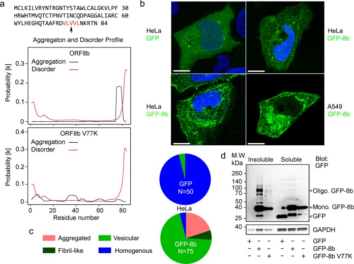
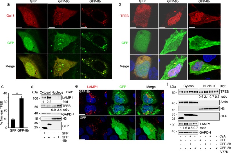
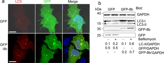
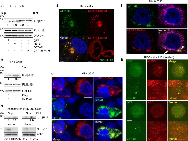
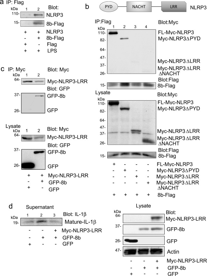
The SARS (severe acute respiratory syndrome) outbreak was caused by a coronavirus (CoV) named the SARS-CoV. SARS pathology is propagated both by direct cytotoxic effects of the virus and aberrant activation of the innate immune response. Here, we identify several mechanisms by which a SARS-CoV open reading frame (ORF) activates intracellular stress pathways and targets the innate immune response. We show that ORF8b forms insoluble intracellular aggregates dependent on a valine at residue 77. Aggregated ORF8b induces endoplasmic reticulum (ER) stress, lysosomal damage, and subsequent activation of the master regulator of the autophagy and lysosome machinery, Transcription factor EB (TFEB). ORF8b causes cell death in epithelial cells, which is partially rescued by reducing its ability to aggregate. In macrophages, ORF8b robustly activates the NLRP3 inflammasome by providing a potent signal 2 required for activation. Mechanistically, ORF8b interacts directly with the Leucine Rich Repeat domain of NLRP3 and localizes with NLRP3 and ASC in cytosolic dot-like structures. ORF8b triggers cell death consistent with pyroptotic cell death in macrophages. While in those cells lacking NLRP3 accumulating ORF8b cytosolic aggregates cause ER stress, mitochondrial dysfunction, and caspase-independent cell death.

Keywords: Cell death and immune response; Inflammasome.


Conflict of interest statement

Conflict of interestThe authors declare that they have no conflict of interest.

Figures

Fig. 1
Fig. 1. ORF8b forms intracellular protein aggregates whose presence depend on V77.
a ORF8b amino acid sequence and predicted aggregation and disorder profile. The amino acid motif predicted to be critical for ORF8b aggregation by PASTA 2.0 (protein aggregation prediction server, http://protein.bio.unipd.it/pasta2/) is colored red. Aggregation and disorder profiles are shown for ORF8b and mutant (V77K) ORF8b. b Confocal microscopy images of HeLa cells transiently transfected with GFP-8b show intracellular patch and fiber-like protein aggregates. Nuclei are counterstained with DAPI and the scale bar is 10 μm. c Quantification of percentage of GFP-positive cells with protein aggregates for GFP and GFP-8b from b. **p < 0.001 with Student’s t-test. N = number of transfected cells examined. d Immunoblots for indicated proteins from triton soluble and triton insoluble fractions of transiently transfected HeLa cells. Experiment repeated a minimum of three times with similar results
Fig. 2
Fig. 2. ORF8b induces ER stress and cell death.
a Confocal microscopy images of HeLa cells transiently transfected with either GFP or GFP-8b and immunostained for the endoplasmic reticulum marker ERp72 (red). Nuclei were counterstained with DAPI and cells were fixed prior to imaging. Individual and merged images are shown, and the scale bar is 5 μm. b Immunoblot looking at the accumulation of the ER stress marker CHOP after transient transfection of GFP, GFP-8b, or GFP-8b V77K in HeLa cells. c Quantification of cell death by Tryptan Blue uptake after overnight transfection of HeLa cells with the indicated proteins. A minimum of three fields with >50 cells per field were quantified. Statistical significance evaluated with Student’s t-test (*p < 0.05, **p < 0.01). Each experiment repeated a minimum of three times
Fig. 3
Fig. 3. ORF8b triggers lysosomal stress and TFEB nuclear translocation.
a, b HeLa cells were transiently transfected with the indicated plasmids. Confocal microscopy images of GFP or GFP-8b co-transfected with RFP-galectin 3 (a) or mCherry-TFEB (b). Individual and merged images are shown, and the scale bar is 5 μm. c The percentage of cells with predominantly nuclear TFEB from part b was quantified for each condition (three fields, minimum of 50 cells/field). **p < 0.01 with Student’s t-test. d Immunoblot to assess LAMP1 and TFEB expression in nuclear and cytosolic fractions separated by SDS-PAGE. HeLa cells were transiently transfected with GFP or GFP-8b. e Representative confocal images from HeLa cell expressing GFP or GFP-8b and immunostained for LAMP1 expression. Individual and merged images are shown, and the scale bar is 5 μm. f Immunoblot of cytosol and nuclear extracts from GFP, GFP-8b, or GFP-8b V77K transfected HeLa cells treated overnight with or without the calcineurin inhibitor cyclosporin A (CsA- 400 nM). Images and blots are representative data from experiments repeated a minimum of 3 times
Fig. 4
Fig. 4. ORF8b enhances autophagic flux and autophagosome formation.
a Confocal microscopy images of HeLa cells co-transfected with the autophagosome marker RFP-LC3 and either GFP or GFP-8b showing GFP-8b induced RFP-LC3 puncta and GFP-8b/RFP-LC3 co-localization. Individual and merged images are shown, and the scale bar is 5 μm. b Immunoblot of LC3 and the indicated proteins from whole cell lysates of GFP or GFP-8b transfected HeLa cells treated with or without Bafilomycin A1 (100 nM, 4 h). Band density quantification was normalized to GAPDH. Images and blots are representative data from experiments repeated a minimum of 3 times
Fig. 5
Fig. 5. ORF8b triggers NLRP3 inflammasome activation.
a Immunoblots for the indicated proteins of PMA differentiated THP-1 cell supernatant and lysates after transient transfection of expression vectors for GFP, 8b-GFP, GFP-8b, or GFP-8b V77K. b Immunoblots for the indicated proteins of PMA differentiated THP-1 cell supernatant and lysates after transient transfection of Flag or 8b-Flag. c Immunoblots for the indicated proteins of HEK 293T cell supernatants and lysates expressing the requisite inflammasome components and either GFP-8b or 8b-Flag with their appropriate control vectors. d Confocal microscopy images of HeLa cells expression NLRP3-DsRed only or NLRP3-DsRed and GFP-8b. Nuclei were counterstained with DAPI and cells were fixed prior to imaging. Individual and merged images are shown, and the scale bar is 5 μm. e Confocal microscopy images of HEK 293T cells expressing NLRP3-DsRed and GFP-8b. Left panels show co-transfected cell and a cell expressing NLRP3-DsRed alone. The right panels show a cell expressing both NLRP3-DsRed and GFP-8b. Individual and merged images are shown, and the scale bar is 5 μm. f Confocal microscopy images of HeLa cells expressing NLRP3-DsRed and GFP-8b and immunostained for TGN38. Individual and merged images are shown, and the scale bar is 3 μm. Images from a single confocal slice. Arrows point areas of overlapping signal. g Confocal microscopy images of THP-1 cells LPS activated and transfected with either GFP or GFP-8b. The following day the cells were immunostained for NLRP3 (upper panels) or ASC (lower panels). Individual and merged images are shown, and the scale bar is 5 μm. Images and blots are representative data from experiments repeated a minimum of 3 times
Fig. 6
Fig. 6. ORF8b interacts with the LRR domain of NLRP3.
a Endogenous NLRP3 immunoblot of Flag immunoprecipitates using cell lysates prepared from THP-1 cells transiently expressing 8b-Flag. The cells were transfected the day prior to use and treated with LPS (50 ng/ml) overnight. b Myc or Flag immunoblot of Flag immunoprecipitates (top) or cell lysates (bottom) prepared from HEK 293 T cells expressing the indicated Myc tagged NLRP3 constructs and 8b-Flag. A schematic representation of the NLRP3 domain organization is shown for clarity. c Myc or GFP immunoblots of Myc immunoprecipitates and cell lysates from HEK 293 cells expressing Myc-NLRP3-LRR and either GFP or GFP-8b. d Indicated immunoblots of cell supernatant and lysates from HEK 293 T cells expressing NLRP3, Casp-1, ASC, full-length IL-1β, and Myc-NLRP3-LRR in the presence of GFP or GFP-8b. Images and blots are representative data from experiments repeated a minimum of 3 times

References

    1. Peiris JS, Guan Y, Yuen KY. Severe acute respiratory syndrome. Nat. Med. 2004;10:S88–S97. doi: 10.1038/nm1143. - DOI - PMC - PubMed
    1. Stadler K, et al. SARS—beginning to understand a new virus. Nat. Rev. Microbiol. 2003;1:209–218. doi: 10.1038/nrmicro775. - DOI - PMC - PubMed
    1. Balboni A, Battilani M, Prosperi S. The SARS-like coronaviruses: the role of bats and evolutionary relationships with SARS coronavirus. New Microbiol. 2012;35:1–16. - PubMed
    1. Zaki AM, van Boheemen S, Bestebroer TM, Osterhaus AD, Fouchier RA. Isolation of a novel coronavirus from a man with pneumonia in Saudi Arabia. N. Engl. J. Med. 2012;367:1814–1820. doi: 10.1056/NEJMoa1211721. - DOI - PubMed
    1. Franks TJ, et al. Lung pathology of severe acute respiratory syndrome (SARS): a study of 8 autopsy cases from Singapore. Hum. Pathol. 2003;34:743–748. doi: 10.1016/S0046-8177(03)00367-8. - DOI - PMC - PubMed