. 2017;4(1):1317690.
Epub 2017 Apr 26.

Developmental exposures to bisphenol S, a BPA replacement, alter estrogen-responsiveness of the female reproductive tract: a pilot study

Affiliations

Developmental exposures to bisphenol S, a BPA replacement, alter estrogen-responsiveness of the female reproductive tract: a pilot study

Corinne E Hill et al. Cogent Med. 2017.

Abstract

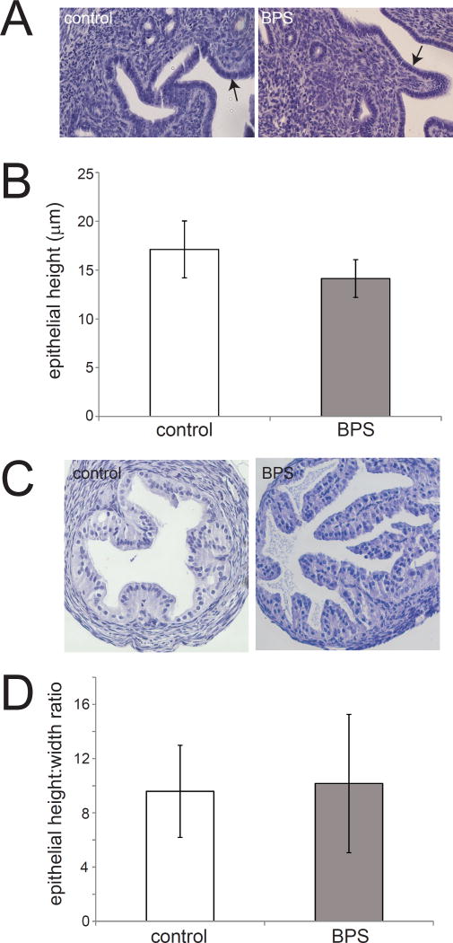
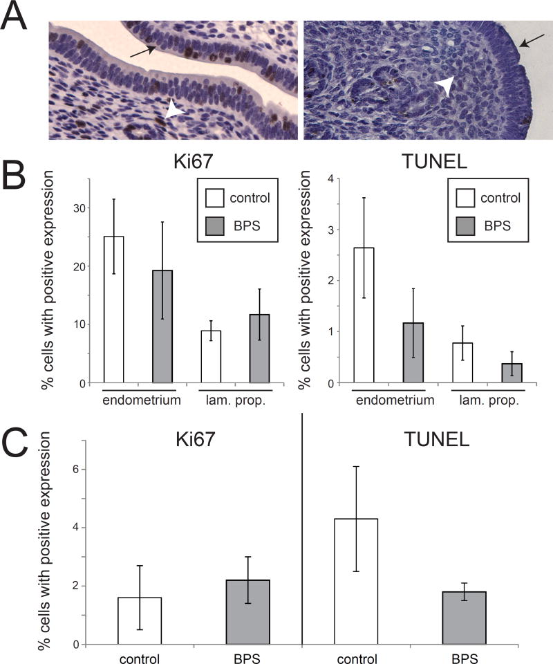
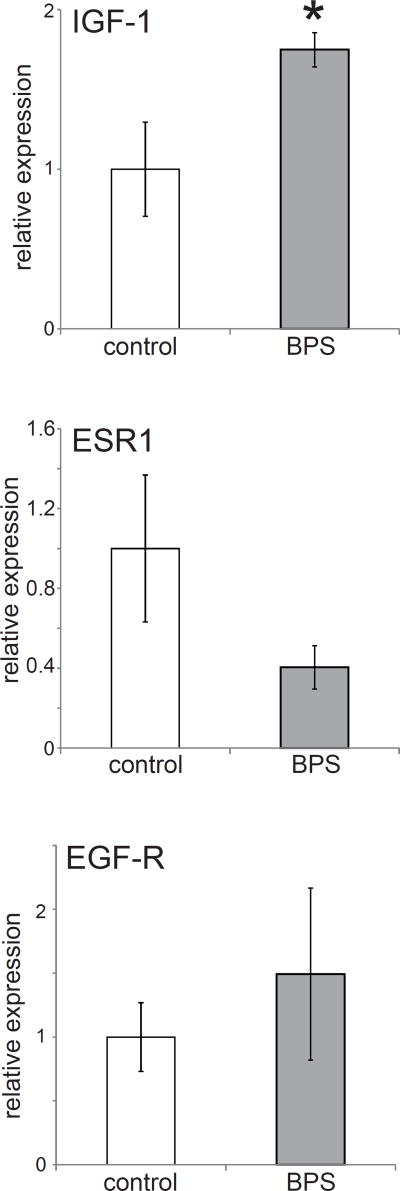
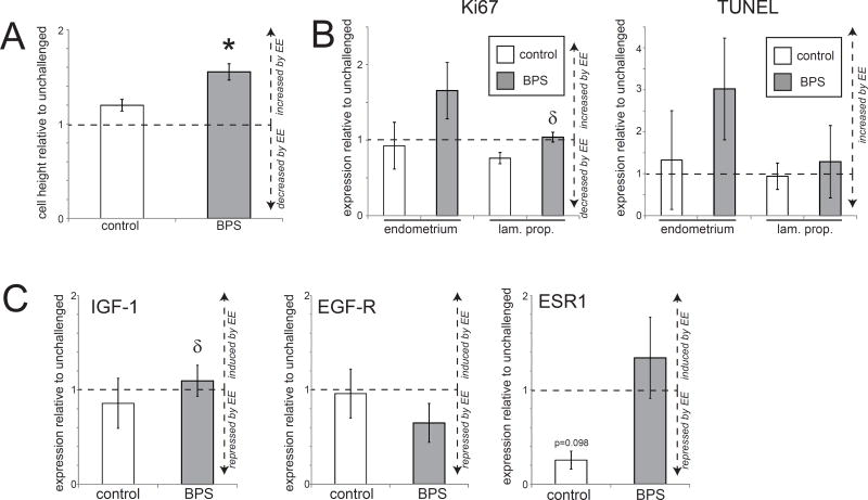
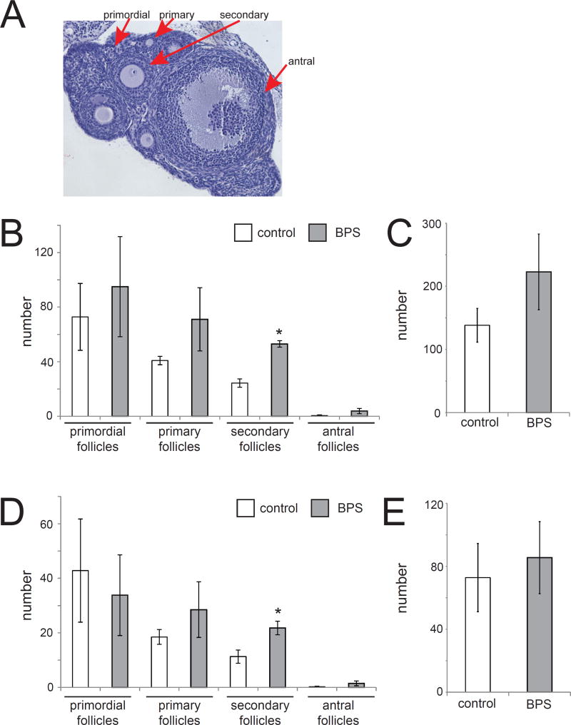
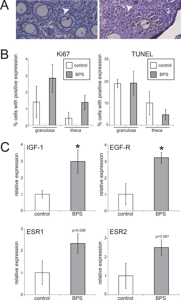
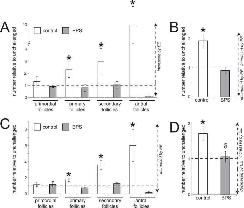
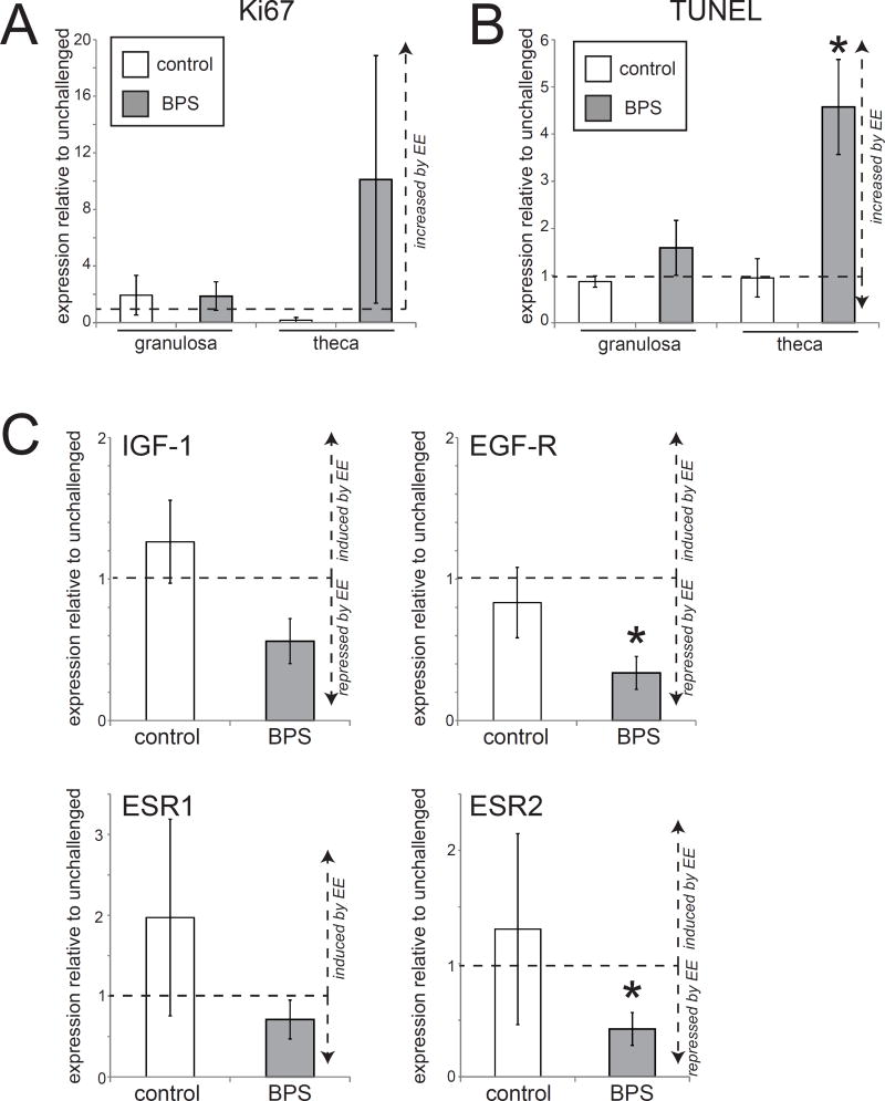
Developmental exposures to bisphenol A (BPA), an estrogen receptor agonist, can disrupt development of the female reproductive tract in rodents and non-human primates. Due to an increased public knowledge of negative health effects associated with BPA exposure, BPA has begun to be phased out of many consumer products and in some cases it has been replaced with structurally similar compounds including bisphenol S (BPS). This study examined CD-1 mice exposed to a low dose of BPS during early development (200 µg/kg/day from gestational day 8 until postnatal day 19). BPS altered expression of estrogen-responsive genes in both the uterus and ovary, and induced increases in ovarian follicular development in pre-pubertal females evaluated at postnatal day 22. Prior studies have revealed that developmental exposures to environmental chemicals including BPA alter the response of animals to hormonal or carcinogen challenges experienced later in life. To evaluate whether early life exposures to BPS alter responses of females to an estrogen challenge, additional females were exposed to ethinyl estradiol from postnatal day 19 through postnatal day 21. BPS-treated females responded abnormally to this estrogen challenge, displaying heightened responses in the uterus and diminished responses in the ovary. Although additional studies are needed to characterize the mechanisms by which BPS alters the female reproductive tract, this pilot study provides evidence that a common BPA replacement chemical may have endocrine disrupting properties.

Keywords: apoptosis; endocrine disruptor; estrogen receptor; ethinyl estradiol; ovarian follicles; proliferation; puberty; uterine endometrium.


Figures

Figure 1
Figure 1. Uterine endometrial cell height and oviduct cell height are not affected by perinatal BPS exposure
A) H&E staining of uterine sections from unexposed and BPS-exposed females. The endometrial layer is indicated by arrows. Photomicrographs were collected with a 20× EpiPlan Objective. B) Quantification of endometrial cell height in unexposed (control) and BPS-exposed females. C) H&E staining of oviduct sections from unexposed and BPS-exposed females. Photomicrographs were collected with a 20× EpiPlan Objective. D) Quantification of oviduct cell height in unexposed (control) and BPS-exposed females. In all groups, n=5 per treatment.
Figure 2
Figure 2. BPS does not alter expression of markers of proliferation or apoptosis in the uterus
A) Expression of Ki67 and TUNEL labeling in the uterus. The endometrial layer is indicated by an arrow and the lamina propria is indicated by an arrowhead. B) Quantification of expression revealed no effect of BPS exposure on Ki67 in either the endometrium or the lamina propria of the uterus. Quantification of TUNEL also showed no effect of BPS on apoptosis in either tissue layer. C) Quantification of Ki67 and TUNEL in the oviduct also revealed no effect of BPS treatment. In all groups, n=5 per treatment.
Figure 3
Figure 3. Developmental exposure to BPS alters gene expression in the female reproductive tract
Expression of three genes in the uterus at PND22. ESR2 expression levels were too low to be accurately quantified in these tissue samples (similar to (Couse et al., 1997)). In all panels, * indicates p<0.05, independent samples t-test. In all groups, n=5 per treatment.
Figure 4
Figure 4. Tissue organization and gene expression in the uterus of BPS-treated females is disrupted by an EE challenge
A) Height of the uterine epithelium was significantly increased in BPS+EE females after an estrogen challenge compared to unchallenged females. In contrast, an estrogen challenge did not significantly affect height of the uterine epithelium in control+EE females. B) Quantification of Ki67 and TUNEL expression in the uterus of control and BPS-treated females after an EE challenge. C) Expression of three genes in uteri is shown relative to unchallenged females from the same perinatal exposure group. Estrogen treatment induced a non-significant decrease in ESR1 in the uterus of control+EE females, but not in BPS+EE females. In all panels, * indicates p<0.05, independent samples t-test comparing EE-challenged with non-challenged females from the same perinatal treatment group (control or BPS). δ indicates significant differences, p<0.05, between control+EE females and BPS+EE females. In all groups, n=5 per treatment.
Figure 5
Figure 5. BPS-induces significant increases in mature ovarian follicles
A) H&E staining of an ovary collected at PND22. Examples of ovarian follicles at various stages of development are indicated. Photomicrographs were collected with a 10× EpiPlan Objective. B) BPS exposure induced significant increases in the number of secondary follicles in the entire ovary. C) The total number of follicles through the entire ovary was increased in BPS-treated females, although this increase was not statistically significant. D) To account for differences in ovarian size, four sections from the middle of the ovary were used to quantify follicle numbers in the various stages of development. BPS induced significant increases in the number of secondary follicles and non-significant increases in the number of primary and antral follicles. E) Non-significant increases were observed in total number of measured in four ovarian sections from BPS-treated ovaries. In all panels, * indicates p<0.05, independent samples t-test. In all groups, n=5 per treatment.
Figure 6
Figure 6. No significant effects of BPS on expression of markers of proliferation or apoptosis in the ovary
A) Expression of Ki67 and TUNEL labeling in the ovary. Granulosa cells are indicated by an arrow and theca cells are indicated by an arrowhead. B) Quantification of expression revealed non-significant increases in Ki67 expression in both cell types in BPS-exposed ovaries. No differences were observed for TUNEL expression in BPS-treated ovaries. Photomicrographs were collected with a 20× EpiPlan Objective. C) Expression of four genes in the ovary at PND22. In all panels, * indicates p<0.05, independent samples t-test. In all groups, n=5 per treatment.
Figure 7
Figure 7. Tissue organization in the ovary of BPS-treated females is disrupted by an EE challenge
A) An estrogen challenge induced significant ovarian development in control+EE females as measured by the increased number of primary, secondary and antral follicles. In contrast, the estrogen challenge had no effect on ovarian development in BPS+EE females. B) Total numbers of follicles counted throughout the entire ovary were significantly increased in control+EE females but unaffected in BPS+EE females. C) Similar patterns were observed when measures of ovarian development were limited to only four sections for specific types of follicles and D) total follicles in four sections of ovary. In all panels, * indicates p<0.05, independent samples t-test comparing EE-challenged with non-challenged females from the same perinatal treatment group (control or BPS). δ indicates significant differences, p<0.05, between EE-challenged controls and EE-challenged BPS-treated females. In all groups, n=5 per treatment.
Figure 8
Figure 8. Significant alterations in apoptosis and gene expression in the ovary of BPS-treated females after an estrogen challenge
A) Quantification of Ki67 in the ovary of control+EE and BPS+EE females after an estrogen challenge. B) Quantification of TUNEL in the ovary of control+EE and BPS+EE females after an estrogen challenge. C) An estrogen challenge decreased the expression of estrogen-sensitive genes in the ovaries of BPS+EE females. In all panels, * indicates p<0.05, independent samples t-test comparing EE-challenged with non-challenged females from the same perinatal treatment group (control or BPS). δ indicates significant differences, p<0.05, between control+EE and BPS+EE females. In all groups, n=5 per treatment.

Similar articles

Cited by

References

    1. Crain DA, Janssen SJ, Edwards TM, et al. Female reproductive disorders: the roles of endocrine-disrupting compounds and developmental timing. Fertil Steril. 2008;90(4):911–40. - PMC - PubMed
    1. Mandrup KR, Jacobsen PR, Isling LK, et al. Effects of perinatal ethinyl estradiol exposure in male and female Wistar rats. Reprod Toxicol. 2013;42:180–91. - PubMed
    1. Soto AM, Rubin BS, Sonnenschein C. Endocrine disruption and the female. In: Gore A, editor. Endocrine-Disrupting Chemicals. Totowa, N.J.: Humana Press; 2007.
    1. McLachlan JA. Commentary: prenatal exposure to diethylstilbestrol (DES): a continuing story. Int J Epidemiol. 2006;35(4):868–70. - PubMed
    1. Herbst AL, Ulfelder H, Poskanzer DC. Adenocarcinoma of the vagina. Association of maternal stilbestrol therapy with tumor appearance in young women. New England Journal of Medicine. 1971;284(15):878–81. - PubMed

LinkOut - more resources