. 2019 Jun 5:2:203.
doi: 10.1038/s42003-019-0442-2. eCollection 2019.

TRAV1-2+ CD8+ T-cells including oligoconal expansions of MAIT cells are enriched in the airways in human tuberculosis

Affiliations

TRAV1-2+ CD8+ T-cells including oligoconal expansions of MAIT cells are enriched in the airways in human tuberculosis

Emily B Wong et al. Commun Biol. .

Abstract

Mucosal-associated invariant T (MAIT) cells typically express a TRAV1-2+ semi-invariant TCRα that enables recognition of bacterial, mycobacterial, and fungal riboflavin metabolites presented by MR1. MAIT cells are associated with immune control of bacterial and mycobacterial infections in murine models. Here, we report that a population of pro-inflammatory TRAV1-2+ CD8+ T cells are present in the airways and lungs of healthy individuals and are enriched in bronchoalveolar fluid of patients with active pulmonary tuberculosis (TB). High-throughput T cell receptor analysis reveals oligoclonal expansions of canonical and donor-unique TRAV1-2+ MAIT-consistent TCRα sequences within this population. Some of these cells demonstrate MR1-restricted mycobacterial reactivity and phenotypes suggestive of MAIT cell identity. These findings demonstrate enrichment of TRAV1-2+ CD8+ T cells with MAIT or MAIT-like features in the airways during active TB and suggest a role for these cells in the human pulmonary immune response to Mycobacterium tuberculosis.

Keywords: Mucosal immunology; T-cell receptor; Tuberculosis.


Conflict of interest statement

Competing interestsThe authors declare no competing interests.

Figures

Fig. 1
Fig. 1
TRAV1-2+ CD8+ T cells from the lung but not the intestine of healthy organ donors respond to mycobacterial infection by producing TNF. a Dot plots showing the frequency of TRAV1-2+ CD8+ T cells among live CD3+ cells in the indicated tissue samples from one donor. b Tissue sections from the 1st and 2nd order bronchi were obtained from healthy individuals (n = 3 biologically independent samples). Immunohistochemistry was performed to quantify CD8+ (median 1.6 × 104 vs. 2 × 104 cells /mm3) and TRAV1-2+ cells (7,000 vs. 4,000 cells/mm3, Supplementary Data). Representative sections from 1st and 2nd order bronchi are depicted (left), showing CD8+ cells (red), TRAV1-2+ cells (green) and cell nuclei (DAPI; blue). c Histograms depicting TNF production (left) and CD161 expression (right) by TRAV1-2+ CD8+ T cells from matched lung parenchyma (green), small intestine lamina propria (LP; blue) and the small intestinal intraepithelial layer (IEL; violet) after overnight stimulation with M. smegmatis-infected antigen-presenting cells (dotted black line indicates the unstimulated control). d Frequency of TRAV1-2+ cells among CD8+ T cells from lung (n = 9 biologically independent samples), mediastinal lymph node (Med LN; n = 11 biologically independent samples), IEL (n = 7 biologically independent samples), LP (n = 8 biologically independent samples), mesenteric lymph node (Mes LN, n = 5 biologically independent samples), and peripheral blood (PBMC; n = 6 biologically independent samples, Supplementary Data). Medians and interquartile ranges are displayed. e Frequency of TNF-producing TRAV1-2+ CD8+ T cells after exposure to M. smegmatis-infected antigen-presenting cells: lung (n = 7 biologically independent samples), Med LN (n = 6 biologically independent samples), IEL (n = 5 biologically independent samples), LP (n = 6 biologically independent samples), Mes LN (n = 2 biologically independent samples), PBMC (n = 12 biologically independent samples, Supplementary Data). From top to bottom, P = 0.035, 0.0025, 0.0023 and 0.0005 (Mann–Whitney U test). Medians and interquartile ranges are displayed
Fig. 2
Fig. 2
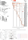
Expansions of MAIT cell-consistent CDR3α‘s are present in tuberculous lung granulomas. a Frequency of TRAV1-2+ sequences as a percentage of all productive TCRα sequences. In some cases, multiple areas of tissue were sampled, ranging from closest (A) to furthest (C) from the site of disease. b MAIT cell TCRα sequences are consistent with similarity scores of 0.95 and 1. Each symbol represents the frequency of TRAV1-2+ or TRAV12-2+ sequences within each similarity score for each donor sample (n = 12 biologically independent samples). c Frequency of total TRAV1-2+ sequences or those with similarity scores of 0.95 and 1 in the lung (n = 3 biologically independent samples) and mediastinal lymph node (LN, 1 sample) from donor 23. Height represents mean, error bars represent standard error. d Frequencies among TRAV1-2+ sequences of the top 10 public and private MAIT cell CDR3α sequences (MAIT Match score ≥0.95) across individual donors and lung samples. e Variation in the number of synonymous nucleotide sequences encoding the five most frequent private (left) and public (right) MAIT cell CDR3α amino acid (aa) sequences from all samples displayed in Fig. 3d. For each aa sequence, each colored bar represents a different nucleotide sequence. The 13 different nucleotide sequences used to generate the shared MAIT cell CDR3α aa sequence CAVLDSNYQLIW are displayed. Text color represents nucleotide origin: purple (TRAV), black (TRAD or n insertion), red (TRAJ). LG lung granuloma, LN lymph node. Source data are provided in Supplementary Data
Fig. 3
Fig. 3
TNF-producing TRAV1-2+ CD8+ cells including oligoclonally expanded MAIT cells are enriched in bronchoalveolar lavage fluid from patients with TB. a Frequency of TRAV1-2+ cells among CD8+ T cells from the bronchoalveolar lavage (BAL) fluid from patients with TB (n = 6 biologically independent samples) and cancer controls (n = 6 biologically independent samples), and among CD8+ T cells in matched peripheral blood samples (PBMC) from patients with TB (n = 5 biologically independent samples) and unmatched peripheral blood samples from healthy controls (n = 13 biologically independent samples). Medians and interquartile ranges are displayed. **P < 0.01; Mann–Whitney U test. b Dot plots showing TNF production by TRAV1-2+ CD8+ T cells in matched BAL and peripheral blood samples (PBMC) from a patient with TB. Cells were stimulated with α−CD2/CD3/CD28 beads. c Frequency of TNF or IL-17 production by TRAV1-2+ CD8+ T cells in matched BAL and peripheral blood samples (PBMC; n = 5 biologically independent samples). Medians and interquartile ranges are displayed. **P < 0.01; Mann–Whitney U test. d Expression of CD161, CD26 and CD103 on TRAV1-2+ CD8+ T cells in matched BAL and peripheral blood samples (PBMC) from patients with TB (n = 4 biologically independent samples). Histograms are mode-normalized. e Frequency of MAIT cell-consistent CDR3α sequences within TRAV1-2+ CD4- T cells in BAL fluid and peripheral blood samples (PBMC) from patients with TB (n = 3 biologically independent samples). Height represents the mean, error bars represent the range. f Relative frequency of CDR3α sequences by MAIT Match Score category in BAL fluid vs. matched peripheral blood (PBMC; n = 3 biologically independent samples). g Depiction of the top 10 most frequent MAIT cell-consistent CDR3α sequences (MAIT Match score ≥0.95) among TRAV1-2+ sequences in each compartment. Legend format: CDR3α aa (# of synonymous nucleotide sequences in peripheral blood, # of synonymous nucleotide sequences in BAL fluid). h IFNγ spot-forming units (SFU) produced by BAL T cell clones stimulated with M. smegmatis-infected or Mtb-infected wildtype (WT) or MR1-KO A549 cells (n=8 biologically independent clones). Height represents the mean of two independent replicates per stimulation, error bars represent the standard deviation. Source data are available in Supplementary Data.
Fig. 4
Fig. 4
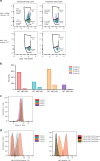
Heterogeneous MR1/5-OP-RU staining of bronchoalveolar TRAV1-2+ CD8+ T cells with MAIT cell-consistent CDR3α‘s and MR1-restricted function. a Frequency of MR1-tetramer+ cells (loaded with active (5-OP-RU) and control (6FP) ligand) in TRAV1-2+ T cells (gated on live, CD3+, CD8+ lymphocytes) from the BAL fluid and peripheral blood of a patient with TB. The proportion of cells utilizing MAIT cell-consistent CDR3α‘s (MAIT Match Score ≥.95) in MR1/5-OP-RU tetramer positive and negative populations are shown. b IFNγ spot-forming units (SFU) produced by four T cell clones generated from BAL fluid and stimulated with M. smegmatis-infected wildtype (WT) or MR1-KO A549 cells, Supplementary Data. c α-TRAV1-2 staining of four T cell clones generated from BAL fluid demonstrates consistent staining. Histograms are mode-normalized. d Binding of MR1/5-OP-RU tetramer on the same four T cell clones generated from BAL fluid demonstrates heterogenous MR1/5-OPRU tetramer staining (left). Binding of MR1/6-FP (control) and MR1/5-OPRU tetramer is shown for two clones (right). Histograms are mode-normalized

References

    1. Porcelli S, Yockey CE, Brenner MB, Balk SP. Analysis of T cell antigen receptor (TCR) expression by human peripheral blood CD4-8- alpha/beta T cells demonstrates preferential use of several V beta genes and an invariant TCR alpha chain. J. Exp. Med. 1993;178:1–16. doi: 10.1084/jem.178.1.1. - DOI - PMC - PubMed
    1. Tilloy F, et al. An invariant T cell receptor alpha chain defines a novel TAP-independent major histocompatibility complex class Ib-restricted alpha/beta T cell subpopulation in mammals. J. Exp. Med. 1999;189:1907–1921. doi: 10.1084/jem.189.12.1907. - DOI - PMC - PubMed
    1. Treiner E, et al. Selection of evolutionarily conserved mucosal-associated invariant T cells by MR1. Nature. 2003;422:164–169. doi: 10.1038/nature01433. - DOI - PubMed
    1. Gold MC, et al. Human mucosal associated invariant T cells detect bacterially infected cells. PLoS Biol. 2010;8:e1000407. doi: 10.1371/journal.pbio.1000407. - DOI - PMC - PubMed
    1. Le Bourhis L, et al. Antimicrobial activity of mucosal-associated invariant T cells. Nat. Immunol. 2010;11:701–708. doi: 10.1038/ni.1890. - DOI - PubMed

Publication types

MeSH terms

Substances